重庆医科大学:《诊断学》课程教学资源(授课教案)19 胸部检查
营庆医科大学脑床半院藏案讲满 重庆医科大学临床学院教案及讲稿 课程名称诊断学 年级2005 授课专业临床医学 教师刘忠 职称教授 授课方式 大课 学时2 题目章节胸部检查 教材名称《诊断学(双语)》 作者吕卓人雷寒 出版社科学出版社 版次第一版 1.familiar with anatomic marks and artificial scribing 教学目的要求 2.grasp the method of inspection,palpation,percussion and auscultation of lung. 3.grasp the clinical meaning of normal breath sound,abnormal breath sound,rales and pleural friction rub. 4.familiar with normal/abnormal respiratory. common classification ofabnormalities of respiration 2. percuss the lower margins of the lung and diaphragmatic movemen the meaning of Rales and rhonchi 1 normal morphology of thoax,common categories of thoracic abnormity and their clinical meaning. 教学重点 common classification of abnormalities of respiratory rhythm and their clinical meaning. correct examination of inspection,palpation,percussion and auscultation clinical meaning of abnormal respiration sound,rales pleural friction rub. English teaching in classes practicing in the hospital and watching videos. 1.kumar &clark.clinical medicine.textbook of medicine,5th edition,edited by Parveen Kumar michael clark.W.B.saunders(elsevier) 2 Davidson.D PcofMe错误链接无效。e2Oth Edition,elsevier company Ltd. 教研 见 制表时间:2005年8月
重庆医科大学临床学院教案讲稿 制表时间:2005 年 8 月 1 重庆医科大学临床学院教案及讲稿 课程名称 诊断学 年级 2005 授课专业 临床医学 教 师 刘忠 职称 教授 授课方式 大课 学时 2 题目章节 胸部检查 教材名称 《诊断学(双语)》 作者 吕卓人 雷寒 出 版 社 科学出版社 版次 第一版 教 学 目 的 要 求 1. familiar with anatomic marks and artificial scribing. 2. grasp the method of inspection, palpation, percussion and auscultation of lung. 3. grasp the clinical meaning of normal breath sound, abnormal breath sound, rales and pleural friction rub. 4. familiar with normal/abnormal respiratory. 教 学 难 点 1. common classification of abnormalities of respiration 2. percuss the lower margins of the lung and diaphragmatic movement 3. the meaning of Rales and rhonchi 教 学 重 点 1 normal morphology of thoax, common categories of thoracic abnormity and their clinical meaning. 2 common classification of abnormalities of respiratory rhythm and their clinical meaning. 3 correct examination of inspection, palpation, percussion and auscultation 4 clinical meaning of abnormal respiration sound, rales pleural friction rub. 外语 要求 English 教学 方法 手段 teaching in classes、practicing in the hospital and watching videos. 参考 资料 1. kumar &clark. clinical medicine. textbook of medicine, 5th edition, edited by Parveen Kumar & michael clark.W.B. saunders(elsevier) company. 2. Davidson. Davidson's Principles and Practice of Medicine. 错误!链接无效。e. 20th Edition, elsevier company Ltd. 教研 室意 见
置庆医科大学床半院藏讲满 讲璃 Physical examination of chest 1.The relation between x-ray study and physical examination of chest Rontgen a Germany physicist discovered the x-ray in 1895,who was awarded the first Nobel for physics in 1901. Since that the X-ray examination has been widely used in clinical especially in diagnosis of pulmonary diseases and it has beer routine tes n every careful study verybody knows In som the ay ink t to a wr ng con n I ca and rery mina of DE detected by x-ray film but some signs like friction nh rales nd whea an he det only by es The relation hetu study and PE is atchin Ty-the picture without sound leaves much to be desired and the sound without the picture may be meaningless.So the x-ray study and PE should supplement,confirm and perfect each other.A right conclusion about the relation between x-ray and PE should like Careful examination of chest should enhance our ability to interpret the x-ray films.and the chest film should serve as a check on the physical examination. normal and artificial landmarks Before amination of the thora rks of the x,let's review that will to he locate physical findings in the chest c We can use thes rtific as a reference to locate the physical findings in th External Landmarks Skeletal landmarks 1.suprasternal noteh (trachea's position) 6.rib 2 manubrium sterni intercostal space(interspace) Sternum (corpus sterni+manubrium sterni 3.sternal angle (Louis angle)a visible scapula the i and serve c angle of the scapula,thehand ihs lie near this noint starting point for counting ribs () at the ba vertebra and posterior ribs 5.xiphoid process (costalvertebral angle) The angle Artificial vertical landmarks 2.midclavicular line 3.anterior posteriormidaxillary line 4.midspinal\vertebral line (posterior midline) 制表时间:2005年8月
重庆医科大学临床学院教案讲稿 制表时间:2005 年 8 月 2 讲 稿 Physical examination of chest 1.The relation between x-ray study and physical examination of chest Rontgen a Germany physicist discovered the x-ray in 1895, who was awarded the first Nobel Prize for physics in 1901. Since that the X-ray examination has been widely used in clinical work especially in diagnosis of pulmonary diseases and it has been included as a routine test in every careful study of patient. Everybody knows in some aspects the X-ray examination is superior to PE and this has led to a wrong conclusion in some doctors’ mind. They think that x-ray films can detect everything that can be found by PE as well as much that cannot be found by PE and rely on x-ray examination that much and so neglect the importance of PE. A lung cancer in early stage possibly can be detected by x-ray film but some signs like friction rub, rales and wheezing can be detected only by our senses. The relation between x-ray study and PE is a little similar to watching TV – the picture without sound leaves much to be desired and the sound without the picture may be meaningless. So the x-ray study and PE should supplement, confirm and perfect each other. A right conclusion about the relation between x-ray and PE should like this: Careful examination of chest should enhance our ability to interpret the x-ray films, and the chest film should serve as a check on the physical examination. 2. normal and artificial landmarks Before we start to discuss the examination of the thorax, let’s review some normal and artificial landmarks of the chest surface that will work as a reference to help us to locate physical findings in the chest correctly. We can use these landmarks (skeletal or artificial lines) as a reference to locate the physical findings in the chest. External Landmarks Skeletal landmarks 1, suprasternal notch (trachea’s position) 6, rib 2, manubrium sterni Sternum (corpus sterni + manubrium sterni) 7, intercostal space (interspace) 3, sternal angle (Louis angle) a visible angulation of the sternum that corresponds to the second rib and serves as a convenient starting point for counting ribs 8, scapula The inferior angle of the scapula, the 7th and 8th ribs lie near this point 4, infrasternal angle (costal angle) epigastric area 9, spinous process The 7th cervical vertebra is usually found with ease at the base of the neck and serves as a convenient landmark to help identify the thoracic vertebra and posterior ribs. 5, xiphoid process 10, costalspinal angle (costalvertebral angle) The angle is 45°normally. Artificial vertical landmarks 1, midsternal line 2, midclavicular line 3, anterior\posterior\midaxillary line 4, midspinal\vertebral line (posterior midline)
露庆医科大学临床半蕊藏案讲满 5.scapular line Natural lacuna and anatomic region 1.axillary fossa 2 suprasternal fossa 3,supraclavicular fossa 4,infraclavicular fossa 5,suprascapular region 6,infrascapular region 7,interscapular region Localizing any findings we should be described as being,(1)so many centimeters medial or lateral to the lines of reference,in (2)a specific interspace or interspaces,for example; 3.Sequence(order)of chest examination There are four procedures(steps)in the chest examination Inspection Palpation Percussion Auscultation p us not to mi spect ol t Xaml on supine position uld ette tera of c nt anging pos Inspection What to observe 1.The general nutrition and musculoskeletal development of the patient (nutritional condition,malnutrition and deformity) 3.The xcessively dry),perspiration(sweat),cyanosis] thorax. e anteroposterior diameter is The out a width of the s ang a 45-deg in atients with emphyse ma the ribs are early horizontal.) The g8retnaction or bule oted is an indication of obstruction to the free inflow of air in the airway Bulging of interspaces may occur in massive pleural effusion,tension pneumothorax asthma and emphysema. 5.The shape and symmetry of chest.Dose the patient have any congenital or secondary abnormality of the chest? Flat chest:APdiameter is decreased and is usually less than half of transverse diameter. Barrel chest:AP diameter is increased and is almost equal to or more than the 制表时间:2005年8月 3
重庆医科大学临床学院教案讲稿 制表时间:2005 年 8 月 3 5, scapular line Natural lacuna and anatomic region 1, axillary fossa 2, suprasternal fossa 3, supraclavicular fossa 4, infraclavicular fossa 5, suprascapular region 6, infrascapular region 7, interscapular region Localizing any findings we should be described as being; (1) so many centimeters medial or lateral to the lines of reference, in (2) a specific interspace or interspaces, for example; “ the point of maximal impulse is located in the left 5th interspace 1cm medial to the midclavicular line.” 3. Sequence(order) of chest examination There are four procedures(steps) in the chest examination Inspection Palpation Percussion Auscultation Experience indicate that following this order when we perform the chest examination will help us not to miss (forget or ignore) any important aspect of the examination. But if the patient is lying down on the bed (in supine position) we would better first examine the anterior and lateral side of chest and then move to the back of patient and this will reduce the number of times the patient changing position Inspection What to observe 1. The general nutrition and musculoskeletal development of the patient (nutritional condition, malnutrition and deformity) 2. The skin and breasts [dehydration (excessively dry), perspiration(sweat), cyanosis] 3. The anteroposterior diameter of the thorax. Normally the anteroposterior diameter is smaller than the transverse diameter. 4. The general slope of the ribs and the width of the subcostal angle. (Normally the ribs are situated at about a 45-degree angle in relation to the spine, but in patients with emphysema the ribs are more nearly horizontal.) 5.The presence of retraction or bulging of interspaces Retraction of interspaces noted on inspiration is an indication of obstruction to the free inflow of air in the airway. Bulging of interspaces may occur in massive pleural effusion, tension pneumothorax asthma and emphysema. 5. The shape and symmetry of chest. Dose the patient have any congenital or secondary abnormality of the chest? Flat chest: AP diameter is decreased and is usually less than half of transverse diameter. Barrel chest: AP diameter is increased and is almost equal to or more than the
重庆医科大半床半院载未讲满 transverse diameter Funnel chest:a depression of the sternum Pigeon breast:an anterior protrusion of the sternum The above all belong to the hypogenesis of thoracic wall(developmental problem) Unilateral retraction of the chest:atelectasis,pleural thickening and trauma The type,rate and depth of quiet breathing riaeabout 16-20 timesinte ands In the mal ratio of 8.Abnormal types of respiration Taehypnea:the breath rate is over 24 times/min Rrad a:the breath rate is below 12 times Apnea:the temporary cessation of breathing Hyp rpnea:an increased depth of breathing (Kussmaul's breathing.is associated with metabolic acidosis) Dyspnea:consciousness of difficulty or effort in breathing Inspiratory dyspnea:effort is the greatest in the inspiratory phase Expiratory dyspnea:effort is the greatest in the expiratory phase Dyspnea Normally a person do es not feel he is taking any effort to breath.Dyspnea describes a condition in which the patient becomes con of difficulty or effort in reathing and has to use greater respiratory effort to satisfy the body's needs. Although dyspnea is an entirely subjective feeling,sever dyspnea can be observed by watching: If the patient is in acute distress? Is there nasal flaring(nose wing is fanning)or pursed lip breathing? Are there audible signs,such as stridor and wheezing? Is cyanosis present? Is the patient using accessory muscles (the muscles of the shoulder and neck)? Will he raise his shoulder and nod his head while he is breathing? All of these signs indicate that the patient is in dyspnea. Inspiratory dyspnea:Tumor,foreign body,severe laryngitis,or extrinsic compression that obstruct trachea or major bronchi and produce an obstruction to the inward flow of air.Inspiratory dyspnea is often accompanied by inspiratory retraction of the interspaces,suprasternal fossa and supraclavicular fossas and an audible stridor if the obstruction located in the trachea or larynx. cle constriction,bronchial inflam and exs ecive secreations in asthma,broncl obstru of the inte 制表时间:2005年8月
重庆医科大学临床学院教案讲稿 制表时间:2005 年 8 月 4 transverse diameter. Funnel chest: a depression of the sternum Pigeon breast: an anterior protrusion of the sternum The above all belong to the hypogenesis of thoracic wall (developmental problem) Unilateral retraction of the chest: atelectasis, pleural thickening and trauma 7. The type, rate and depth of quiet breathing Normal respiration In the adult at rest the normal respiratory rate is about 16-20 times a minute and is quite regular in depth and rhythm. The normal ratio of respiratory rate to pulse rate is approximately 1 : 4 and the normal ratio of inspiration to expiration is 1 ~ 2.5 8.Abnormal types of respiration Tachypnea: the breath rate is over 24 times/min Bradypnea: the breath rate is below 12 times/min Apnea: the temporary cessation of breathing Hyperpnea: an increased depth of breathing (Kussmaul’s breathing, is associated with metabolic acidosis) Dyspnea: consciousness of difficulty or effort in breathing Inspiratory dyspnea: effort is the greatest in the inspiratory phase Expiratory dyspnea: effort is the greatest in the expiratory phase Dyspnea Normally a person does not feel he is taking any effort to breath. Dyspnea describes a condition in which the patient becomes conscious of difficulty or effort in breathing and has to use greater respiratory effort to satisfy the body’s needs. Although dyspnea is an entirely subjective feeling, sever dyspnea can be observed by watching: If the patient is in acute distress? Is there nasal flaring(nose wing is fanning) or pursed lip breathing? Are there audible signs, such as stridor and wheezing? Is cyanosis present? Is the patient using accessory muscles (the muscles of the shoulder and neck)? Will he raise his shoulder and nod his head while he is breathing? All of these signs indicate that the patient is in dyspnea. Inspiratory dyspnea: Tumor, foreign body, severe laryngitis 喉 炎 , or extrinsic compression that obstruct trachea or major bronchi and produce an obstruction to the inward flow of air. Inspiratory dyspnea is often accompanied by inspiratory retraction of the interspaces, suprasternal fossa and supraclavicular fossas and an audible stridor if the obstruction located in the trachea or larynx. Expiratory dyspnea: obstruction in the bronchioles and smaller bronchi because of bronchial smooth muscle constriction, bronchial inflammation and exssecive secreations, as in asthma, bronchitis, and obstructive emphysema. Expiration is prolonged because of the obstruction to the outflow of air. On the contrary to the inspiratory dyspnea, it may be accompanied by bulging of the interspaces

露庆医科大学临床半蕊藏案讲满 Pusing our hands to touch patient and it may or supplement the findings already noted during inspection Palpation is the process of touching the patient in an effort to evaluate (1)cervical and axillary lymph nodes (2)trachea's position(position of the mediastinum) (3)thoracic expansion tenderness (5)any chest abnormalities that can not be seen (6)tactile fremitus (vocal fremitus) The term fremitus means vibration and tactile means touch(feeling) The sound that arise in the larynx are transmitted down along the bronchial system into each lung setting the thoracic wall in motion. Thus vibration are produced in the chest wall that can be felt by the hand I of the examiner.To elicit vocal fremitus the patient is with an unifor compa tr on c remit areas of th o fe yo place the corresponding areas so you can compare th Normal variations of fremitus Normally thereare several factorswill infuence the intensity of fremitu duce mo inte ice low nitch ofthe 3)N thickn ss of the ore intens than in obe atient (4)varying relations of the bronehi to the chest wall In general,vocal fremitus is most intense in the regions of the thorax where the large bronchi are the closest to the chest wall It should be pointed out that only big changes in the lung give palpable fremitus and so we shouldn't expect that small patches of early tuberculosis or pneumonia would be accompanied by significant changes of the vocal fremitus. Abnormal variations offremitus Decreased or absent tactile vocal fremitus excess air in the lung (emphysema)because air transmits vibrations are less intense than the tissue. obstruction of major bronchus (obstructive atelectasis)When a tumor occlude the bronchus the transmission of vibration will be diminished othorax the e prne mthe (3)subcutaneous emphysema (the escape of air from the lungs or outside into the subcutaneous tissues as the result of injuries to or operation on the thorax. Increased tactile vocal fremitus 制表时间.2005年8月
重庆医科大学临床学院教案讲稿 制表时间:2005 年 8 月 5 Palpation using our hands to touch patient and it may confirm or supplement the findings already noted during inspection Palpation is the process of touching the patient in an effort to evaluate ⑴ cervical and axillary lymph nodes ⑵ trachea’s position (position of the mediastinum) ⑶ thoracic expansion ⑷ tenderness ⑸ any chest abnormalities that can not be seen ⑹ tactile fremitus (vocal fremitus) The term fremitus means vibration and tactile means touch(feeling) The sound that arise in the larynx are transmitted down along the bronchial system into each lung setting the thoracic wall in motion. Thus vibration are produced in the chest wall that can be felt by the hand of the examiner. To elicit vocal fremitus the patient is asked to say “e-e-e” with an uniform(constant) intensity voice so that the examiner can better compare the transmission of the fremitus in different areas of the chest. To feel the vocal fremitus you must place the palmar side of your fingers against the chest wall in corresponding areas so you can compare the two sides at the time. Normal variations of fremitus Normally there are several factors will influence the intensity of fremitus (1)intensity of the voice Loud and deep voice produce more intense fremitus (2)pitch of the voice low pitch of the voice produce more strong fremitus (3)varying thickness of the thoracic wall In a thin person the vibrations will be more intense than in obese patient. (4)varying relations of the bronchi to the chest wall In general, vocal fremitus is most intense in the regions of the thorax where the large bronchi are the closest to the chest wall ※ It should be pointed out that only big changes in the lung give palpable fremitus and so we shouldn’t expect that small patches of early tuberculosis or pneumonia would be accompanied by significant changes of the vocal fremitus. Abnormal variations of fremitus Decreased or absent tactile vocal fremitus excess air in the lung (emphysema) because air transmits vibrations are less intense than the tissue. obstruction of major bronchus (obstructive atelectasis) When a tumor occlude the bronchus the transmission of vibration will be diminished. (1) fibrous thickening of the pleura (pleurisy, empyema) (2) pleural effusion or pneumothorax the accumulation of fluid or air in the pleural space that will definitely decrease the transmission of vibration (3) subcutaneous emphysema (the escape of air from the lungs or outside into the subcutaneous tissues as the result of injuries to or operation on the thorax. Increased tactile vocal fremitus
露庆医科大半脑床半院教未讲满 Consolidation of lungs (lobar pneumonia,pulmonary infarction).Because solid materials transmit vibrations more readily than air-filled spaces do.In pneumonia the alveoli are consolidated because of filled with inflammatory cells and red blood cells. Percussion Percussion refers to the tapping on a surface to determine the underlying structure 1.Percussion methods Fordelepeg (1)Med ssion(fir nthe border (2)Immediate percussion(direct percussion) For detecting gross changes like pleural effusion and pneumothorax 2.Precautions of percussion If you want to get more information from percussion you should pay attention to the following notes. (1)The distal phalanx of the pleximeter finger must be pressed firmly on the chest wall. (2)The plexor finger should strike the pleximeter finger only instantaneously and must be immediately withdrawn one side of the thorax with the opposite side as he proce compare (5)As the ex ner listens to the sound elicited by be made ofall the chrco tetoefomed cussion,a careful analysis should 3. Percussion sounds (1)The character of percussion notes are dependent upon the air-tissue ratio they have. Resonance Hyperresonance or Tympany (sound of striking drum) Dullness(flatness) Percussion Source Pitch Quality Duration Resonance Normal lung Long Hyperresonance Empnysema Louder/lowe Longer Tympany 制表时间:2005年8月
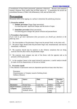
重庆医科大学临床学院教案讲稿 制表时间:2005 年 8 月 6 Consolidation of lungs (lobar pneumonia, pulmonary infarction). Because solid materials transmit vibrations more readily than air-filled spaces do. In pneumonia the alveoli are consolidated because of filled with inflammatory cells and red blood cells. Percussion Percussion refers to the tapping on a surface to determine the underlying structure. 1. Percussion methods ⑴ Mediate percussion (finger-finger percussion) For detecting small, superficial changes and the border of organ ⑵ Immediate percussion (direct percussion) For detecting gross changes like pleural effusion and pneumothorax 2. Precautions of percussion If you want to get more information from percussion you should pay attention to the following notes. ⑴ The distal phalanx of the pleximeter finger must be pressed firmly on the chest wall。 ⑵ The plexor finger should strike the pleximeter finger only instantaneously and must be immediately withdrawn ⑶ The examiner should also be sensitive to the vibratory sensations that are being received from the chest wall by the pleximeter finger. ⑷ The examiner must compare one side of the thorax with the opposite side as he proceeds with the percussion. ⑸ As the examiner listens to the sound elicited by percussion, a careful analysis should be made of all the characteristics of the tone so formed 3. Percussion sounds ⑴The character of percussion notes are dependent upon the air-tissue ratio they have. Resonance Hyperresonance or Tympany (sound of striking drum) Dullness (flatness) Percussion Source Pitch Quality Duration The time sustained Resonance Normal lung Loud / low Hollow Long Hyperresonance Tympany Emphysema Pneumothorax Stomach, bowel Louder / lower Hollow, Striking a empty barrel Longer
露庆医科大学临床半蕊裁案讲满 Dullness High Dull short Striking a resonance and Atelectasis barrel huperresonance Consolidation filled with Liver,heart water (2)Path of percussion (3)Percuss the lower margins of the lung Done in the midclavicular lines,midaxillary lines and scapular lines of both sides. Patient breath quietly,percuss from a resonant area down to a dull point. (4)Percuss to detect diaphragmatic movement at scapular lines Auscultation Auscultation is the technique of listening to sounds produced in the body. Auscultation of the chest is used to identify lung sounds and can yield information about the flow of gas through the tracheobronchial tree and the presence of alveolar consolidation,secretions,or bronchial obstruction. 1.The stethoscope usually has two heads The bell is used to detect low-pitched sounds(the murmur of mitral stenosis and must be loosely applied to the skin;if it is pressed too tightly.the skin will act as a diaphragm and the lower-pitched sounds will be filtered out. The diaphragm is used to detect high-pitched sounds and is applied firmly to the skin Because most breath sounds are high pitched.the diaphragm of the stethoscope is used toevaluate lung sounds. 2.Normal breath sounds Breath sounds Pitch Quality Area Ratio ofI/E Normal Loud and tubular Trachea and over stemum bronchial high breath sounds Bronchovesicu Mixture Mixture Each side of the sternum in lar sounds the first and second nterspaces scapulae Soft and low Sighing Over the most areas of lung 制表时间:2005年8月
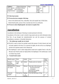
重庆医科大学临床学院教案讲稿 制表时间:2005 年 8 月 7 Dullness The opposite of resonance and huperresonance Pleural thickening Pleural effusion Atelectasis Consolidation Liver, heart High Dull Striking a barrel filled with water short ⑵ Path of percussion ⑶ Percuss the lower margins of the lung Done in the midclavicular lines, midaxillary lines and scapular lines of both sides. Patient breath quietly, percuss from a resonant area down to a dull point. ⑷ Percuss to detect diaphragmatic movement at scapular lines Auscultation Auscultation is the technique of listening to sounds produced in the body. Auscultation of the chest is used to identify lung sounds and can yield information about the flow of gas through the tracheobronchial tree and the presence of alveolar consolidation, secretions, or bronchial obstruction. 1. The stethoscope usually has two heads ★ The bell is used to detect low-pitched sounds (the murmur of mitral stenosis and must be loosely applied to the skin; if it is pressed too tightly, the skin will act as a diaphragm and the lower-pitched sounds will be filtered out. ★ The diaphragm is used to detect high-pitched sounds and is applied firmly to the skin. Because most breath sounds are high pitched, the diaphragm of the stethoscope is used to evaluate lung sounds. 2. Normal breath sounds Breath sounds Pitch Quality Area Ratio of I/E Normal bronchial breath sounds Loud and high tubular Trachea and over sternum Bronchovesicu lar sounds Mixture Mixture Each side of the sternum in the first and second interspaces Between the scapulae Over the apices Vesicular sounds Soft and low Sighing or rustling Over the most areas of lung
重庆医科大半床半院载未讲满 In the normal lung,the sounds heard on auscultation are the sounds of air rushing through the tubeyoke structures of the tracheobronchial tree.Since the rate of flow changes very markedly between the trachea and the alveoli,the nature and pitch of the breath sounds change also. 3.Abnormal breath sounds Bronchial breath sounds-its presence over the lungs always indicates disease (consolidation) Decreased or absent breath sounds Decreased breath sounds(distant breath sounds) (1)emphysema (2)pleural thickening (3)restriction of thoracic excursion ◇pleurisy ◇rib fracture ascites,pregnancy,post operation of abdomen Absent breath sounds (1)pleural effusion (②)pneumothorax (3)atelectasis肺不张(complete bronchial obstruction) Rales and rhonchi (adventitious sounds DRales (bubble.crackles,rattling)-a shower of discrete sounds resulting from the passage of air through secretions in the respiratory tract and from reinflation of the alveol nd bronchioles,the walls of which have become adherent as result of moisture. Fine rales(crepitus) ◇Medium rales ◇Coarse rales Rales Quality/Pitch Origin Time of After Fine Alveoli of Not cleared High Mediu Medium Bronchiol Middle Not Inflammation of m es cleared smaller bronchi 制表时间:2005年8月
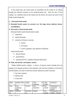
重庆医科大学临床学院教案讲稿 制表时间:2005 年 8 月 8 In the normal lung, the sounds heard on auscultation are the sounds of air rushing through the tubeyoke structures of the tracheobronchial tree. Since the rate of flow changes very markedly between the trachea and the alveoli, the nature and pitch of the breath sounds change also. 3. Abnormal breath sounds ★ Bronchial breath sounds—its presence over the lungs always indicates disease (consolidation) ★ Decreased or absent breath sounds Decreased breath sounds (distant breath sounds) ⑴ emphysema ⑵ pleural thickening ⑶ restriction of thoracic excursion ◇ pleurisy ◇ rib fracture ◇ ascites, pregnancy, post operation of abdomen Absent breath sounds ⑴ pleural effusion ⑵ pneumothorax ⑶ atelectasis 肺不张 (complete bronchial obstruction) ★ Rales and rhonchi ( adventitious sounds ) ①Rales (bubble,crackles, rattling) - a shower of discrete sounds resulting from the passage of air through secretions in the respiratory tract and from reinflation of the alveoli and bronchioles, the walls of which have become adherent as result of moisture. ◇ Fine rales (crepitus) ◇ Medium rales ◇ Coarse rales Rales Quality/ Pitch Origin Time of occurring After cough Indication Fine Fine, crackling/ High Alveoli End of inspiration Not cleared Inflammation or congestion of alveoli Mediu m Medium Bronchiol es Middle Not cleared Inflammation of smaller bronchi
露庆医科大学临床半蕊裁案讲满 or bronchioles Coarse Bubbline/ Bronchi Early Cleared Inflammation or secretion of Low bronch and trachea 2Rhonchi-a continuous sounds produced by the passage of air through the narrowed rachea,bronchi or bronchioles,heard in expiratory phase Wheezing (sibilant):high pitched sound caused by air flow through constricted airways,m often he ◇Sonorous. in expiratory ph roughened inflamed surface of pleura rubbing togethe during respiration producesa very characteristic soun Quality Area being Time of occurring Factors of influence heard Pleural I Coarse Lower part of At the end of Taking deep breath friction Creaking the chest in the inspiration or the Firmly pressing the rub Grating midaxillary beginning of stethoscope over the expiration thoracic wall 3Vocal resonance:the spoken voice as heard over the normal lung. It varies in exactly the same fashion as does vocal fremitus but is more sensitive Bronchophony-the sound becomes louder,clearer and even the pronunciation of words he heard。 Pectoriloquy-much louder than normal,pronuciation of the words can be heard clearly. ◇Egophony-nature of the sound changes.When patient says“E"it sounds like“A"。 Whispered pectoriloquy-ask patient to whisper"1.2.3"In a normal patient,the sound is very weak. In a patient with lung consolidation,it sounds very clear.and high-pitched. Documenting auscultation findings EXAMPLE: Late inspiratory fine crackles were heard over the right base posteriorly along the midscapular line,and coarse crackles were heard throughout inspiration and expiration over the left apex anteriorly near the midclavicular line. 22 Whatthe correct order of physical eamination? 思考 t's ir 5 Which dis 6 What will affect the gauon2 of the chest? 7 What's the relationship between the amount of pleural effusion and its ntoms? 8 What are abnormal types of respiration? 制表时间:2005年8月
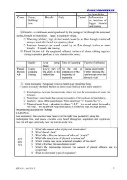
重庆医科大学临床学院教案讲稿 制表时间:2005 年 8 月 9 or bronchioles Coarse Coarse, Bubbling/ Low Bronchi Early Cleared Inflammation or secretion of bigger bronchi and trachea ②Rhonchi - a continuous sounds produced by the passage of air through the narrowed trachea, bronchi or bronchioles,heard in expiratory phase. ◇ Wheezing (sibilant): high pitched sound caused by air flow through constricted airways, most often heard in expiratory phase. ◇ Sonorous: lower-pitched sound caused by air flow through trachea or main bronchi. It sounds like snoring. ③ Pleural friction rub: the roughened inflamed surfaces of pleura rubbing together during respiration produces a very characteristic sound. Quality Area being heard Time of occurring Factors of influence Pleural friction rub Coarse Creaking Grating Lower part of the chest in the midaxillary At the end of inspiration or the beginning of expiration Taking deep breath Firmly pressing the stethoscope over the thoracic wall ③ Vocal resonance: the spoken voice as heard over the normal lung. It varies in exactly the same fashion as does vocal fremitus but is more sensitive ◇ Bronchophony– the sound becomes louder, clearer and even the pronunciation of words can be heard。 ◇ Pectoriloquy– much louder than normal, pronuciation of the words can be heard clearly。 ◇ Egophony– nature of the sound changes. When patient says “E” it sounds like “A”。 ◇ Whispered pectoriloquy – ask patient to whisper “1,2,3”. In a normal patient, the sound is very weak. In a patient with lung consolidation, it sounds very clear, and high-pitched。 Documenting auscultation findings EXAMPLE: Late inspiratory fine crackles were heard over the right base posteriorly along the midscapular line, and coarse crackles were heard throughout inspiration and expiration over the left apex anteriorly near the midclavicular line。 思考 题及 预习 1 What’s the correct order of physical examination? 2 What’s barrel chest? 3 What’s the difference between of rales and rhonchi? 4 What’s the importance of physical examination? 5 Which disease may cause unilateral retraction of the chest? 6 What will affect the auscultation result? 7 What’s the relationship between the amount of pleural effusion and its symptoms? 8 What are abnormal types of respiration?
重庆医科大半临床半院载案讲满 教案讲稿质量评价表 B D 权重 评估内容 权重 较好 一船 1.0-0.90.89 0.79- 0.59-0 编写认真、教学态度端正 教学目的明确、概念清楚、内容准确 20 3.教学注意系统性及先进性 15 4. 重点突出、难点清楚 15 100 5. 教学方法、手段适当 10 6. 运用专业外语适当、准确 10 1. 理论联系实际、举例恰当 10 8.知识容量密度适宜、时间分配合理 评价得分 (A级=100-90分:B级=89-80分:C级=79-60分:D级=59-0分 意见 评价者: 评价时间: 制表时间:2005年8月 10
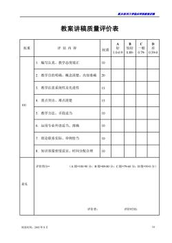
重庆医科大学临床学院教案讲稿 制表时间:2005 年 8 月 10 教案讲稿质量评价表 权重 评 估 内 容 权重 A 好 1.0-0.9 B 较好 0.89- C 一般 0.79- D 差 0.59-0 100 1. 编写认真、教学态度端正 10 2. 教学目的明确、概念清楚、内容准确 20 3. 教学注意系统性及先进性 15 4. 重点突出、难点清楚 15 5. 教学方法、手段适当 10 6. 运用专业外语适当、准确 10 7. 理论联系实际、举例恰当 10 8. 知识容量密度适宜、时间分配合理 10 意见 评价得分= (A 级=100-90 分;B 级=89-80 分;C 级=79-60 分;D 级=59-0 分) 评价者: 评价时间:
按次数下载不扣除下载券;
注册用户24小时内重复下载只扣除一次;
顺序:VIP每日次数-->可用次数-->下载券;
- 重庆医科大学:《诊断学》课程教学资源(授课教案)10 发热.doc
- 重庆医科大学:《诊断学》课程教学资源(授课教案)12 血管检查.doc
- 重庆医科大学:《诊断学》课程教学资源(授课教案)15 超声心动图检查.doc
- 重庆医科大学:《诊断学》课程教学资源(授课教案)13 循环系统主要症状与体征.doc
- 重庆医科大学:《诊断学》课程教学资源(授课教案)20 呼吸系统症状和体征.doc
- 重庆医科大学:《诊断学》课程教学资源(授课教案)11 心脏检查.doc
- 重庆医科大学:《诊断学》课程教学资源(授课教案)14 心电图.doc
- 重庆医科大学:《诊断学》课程教学资源(授课教案)06 腹泻.doc
- 重庆医科大学:《诊断学》课程教学资源(授课教案)05 腹痛.doc
- 重庆医科大学:《诊断学》课程教学资源(授课教案)07 黄疸.doc
- 重庆医科大学:《诊断学》课程教学资源(授课教案)02 水肿.doc
- 重庆医科大学:《诊断学》课程教学资源(授课教案)01 绪论与问诊.doc
- 重庆医科大学:《诊断学》课程教学资源(授课教案)08 腹部检查.doc
- 重庆医科大学:《诊断学》课程教学资源(授课教案)09 病历与诊断.doc
- 重庆医科大学:《诊断学》课程教学资源(授课教案)03 呕血.doc
- 重庆医科大学:《诊断学》课程教学资源(授课教案)04 便血.doc
- 重庆医科大学:《实验诊断学》课程教学资源(PPT课件)第八讲 脑脊液常规及生殖系统检查.ppt
- 重庆医科大学:《实验诊断学》课程教学资源(PPT课件)第七讲 大便常规、免疫学检查及心肌标志物检查.ppt
- 重庆医科大学:《实验诊断学》课程教学资源(PPT课件)第六讲 肾功能检查(主讲:唐敏).ppt
- 重庆医科大学:《实验诊断学》课程教学资源(PPT课件)第四讲 血栓与出血检查(主讲:胥文春).ppt
- 重庆医科大学:《诊断学》课程教学资源(授课教案)18 呼吸困难.doc
- 重庆医科大学:《诊断学》课程教学资源(授课教案)16 咳嗽、咳痰.doc
- 重庆医科大学:《诊断学》课程教学资源(授课教案)17 咯血.doc
- 医学临床本科《MRI诊断学》课程教学大纲 Magnetic Resonance Imaging Diagnosis.doc
- 医学临床本科《影像诊断学》课程教学大纲 medical imaging(包含MRI部分).doc
- 医学影像本科《MRI诊断学》课程教学大纲(Magnetic Resonance Imaging Diagnosis).doc
- 石河子大学:《MRI诊断学》课程教学资源(讲稿,共五章).doc
- 石河子大学:《MRI诊断学》课程教学课件(PPT讲稿)第一章 MRI总论.ppt
- 石河子大学:《MRI诊断学》课程教学课件(PPT讲稿)第三章 呼吸系统 3.1 3.2 正常及异常.ppt
- 石河子大学:《MRI诊断学》课程教学课件(PPT讲稿)第三章 呼吸系统 3.3 支气管扩张及肺部炎症.ppt
- 石河子大学:《MRI诊断学》课程教学课件(PPT讲稿)第三章 呼吸系统 3.4 肺结核.ppt
- 石河子大学:《MRI诊断学》课程教学课件(PPT讲稿)第三章 呼吸系统 3.5 肺肿瘤.ppt
- 石河子大学:《MRI诊断学》课程教学课件(PPT讲稿)第三章 呼吸系统 3.6 纵膈疾病.ppt
- 石河子大学:《MRI诊断学》课程教学课件(PPT讲稿)第四章 循环系统(冠脉及主动脉).ppt
- 石河子大学:《MRI诊断学》课程教学课件(PPT讲稿)第五章 消化系统和腹膜腔 5.2 急腹症.ppt
- 石河子大学:《MRI诊断学》课程教学课件(PPT讲稿)第五章 消化系统和腹膜腔 5.3 肝脏.ppt
- 石河子大学:《MRI诊断学》课程教学课件(PPT讲稿)第五章 消化系统和腹膜腔 5.4 胆道系统疾病.ppt
- 石河子大学:《MRI诊断学》课程教学课件(PPT讲稿)第五章 消化系统和腹膜腔 5.5 胰腺.ppt
- 石河子大学:《MRI诊断学》课程教学课件(PPT讲稿)第五章 消化系统和腹膜腔 5.6 脾脏.ppt
- 石河子大学:《MRI诊断学》课程教学课件(PPT讲稿)第五章 消化系统和腹膜腔 5.1 食管及胃肠道疾病的CT诊断.ppt
