重庆医科大学:《诊断学》课程教学资源(授课教案)14 心电图
重庆医科大学脑床学院载未讲满 重庆医科大学临床学院教案及讲稿 (教案) 课程名称诊断学 年级2005级 授课专业临床医学 教师雷寒 职称教授 授课方式大课 学时6 题目章节心电图检查 教材名称诊断学 作者吕卓人雷寒 出版社科学出版社 版次第一版 目的要 了解心电图的重要性、临床价值、心电图的基本知识、正常心电图的特点、各 难 教学 常见心律素乱、常见心律紊乱的心电图特点(三种早搏、心房颜动、房、结、室心动过速、时 颜)传导阻滞、心肌梗塞的演变、心肌梗塞的心电图分类。 ECG characteris 点 r conduction bloEGchamgenartionE classification of myocardial infarction 外语 掌握基本专业术语 教学 多媒体图形+文字+讲解(mutimedia+text+-lecture) 各版的诊断学,比如此6版P534,图5一1一46可以认为是错误的,必须给学生说明 such as 6 edition page 534 picture 5-1-46 is not correct and should b 教研 见 制表时间:2005年9月
重庆医科大学临床学院教案讲稿 制表时间:2005 年 9 月 1 重庆医科大学临床学院教案及讲稿 ( 教 案 ) 课程名称 诊断学 年级 2005 级 授课专业 临床医学 教 师 雷寒 职称 教授 授课方式 大课 学时 6 题目章节 心电图检查 教材名称 诊断学 作者 吕卓人 雷寒 出 版 社 科学出版社 版次 第一版 教 学 目 的 要 求 诊断学阶段、了解心电图的重要性、临床价值、心电图的基本知识、正常心电图的特点、各种 心律紊乱的心电图特点等。(Understand the importance, clinical value, and basic knowledge of ECG. Understand the characteristic of normal ECG, arrhythmia, etc) 教 学 难 点 具体的数据和心电图特点、常见心律紊乱心电图的特点。(Data and characteristic of ECG, ECG characteristic of common found arrhythmia) 教 学 重 点 常见心律紊乱、常见心律紊乱的心电图特点(三种早搏、心房颤动、房、结、室心动过速、时 颤)传导阻滞、心肌梗塞的演变、心肌梗塞的心电图分类。(Common arrhythmia, ECG characteristic of common arrhythmia (Three types premature complex, Atrial fibrillation, Atrial, Junctional, ventricle tachycardia, ventricle fibrillation), conduction block, ECG change of myocardial infarction, ECG classification of myocardial infarction 外语 要求 掌握基本专业术语 教学 方法 手段 多媒体图形+文字+讲解 (multimedia+text+lecture) 参考 资料 各版的诊断学,比如此 6 版 P534,图 5-1-46 可以认为是错误的,必须给学生说明。 Diagnostics textbook, such as 6th edition page 534 picture 5-1-46 is not correct and should be explained to student 教研 室意 见
营庆医科大半脑床半院载来沙满 (讲稿) 教学内容 铺助毛段 时间分配 心电图 【概述】(Basic Theory) 心脏机械活动前,所产生的综合电活动,用仪器记录下来,称心 第1学时 (By using device to record the electrical activity of the heart which generates ach time (图形、文字、讲解 ),不用EKG((don'useUKG) First teaching hou (multimediatex 临床价信aa: 心律紊乱(A -pen type. -matrix type) cture) 肥大Atri t-but can'testablish the etiology of the diagnosis) 传导系统(Conduction system):窦房结(Sinoatrial node)一结间束Internodal pathways) 一房室结(Atrioventricular node) 希氏束(His bundle)→左、右束支(Left and right bundle branch)→浦倾野氏纤维(Purkinje fiber)。 【心电导联】(E1e 联6见书508页,图5肢体联邮 前导联(S eads and。 510 5- -12fo dial ight of ster num) -位于胸骨左缘第4肋间。(4"intercostals space,left of sterne m) 一位于V和V,之间的中点。(midway between V2andV,) -位于左锁骨中线与第5肋间相交处。(5 intercostals space in midclavicular line) 一位于腋前线V水平处。 (same level as V,at anterior axillary line) 位于左腋中线V,水平处。(same level as in the midaxillary line 落用为g导联(The nine ost common1 eds used))、I、avR、avL、aF、 Infarc tion):加作2V,V,共计l2导联。(Make also Va,V,V,hic ant 、每小格(Every1 small box):横向表示时间为0.04S(horizontal indicate time,which is0.04s):纵向(电压)为0.1mW(/m)(vertical(voltage)is 0.1mN(/mm)) 说明(Note):上述一般情况下,走低速度为25/S。(The above explanation is valid if the ECG recorded at the speed of 25 mm/s) 二、心率测定:(Heart rate calculation) 1、60/R-R间期(60/R-R interval),如(for example))R-R=0.8S,HR=60/0.8 2、 calcul ated by)155-(R-R间期)(interval):15 d on the he from 60 to 100 time 3、数三个大格的R波数×20。(Calculate the number of R wave in3larg hoxes and then x20) 【心电轴】临床价值不大,看书、目测法:([Axis],clinically doesn'thav significant value,look at the book how to determine axis by a glance) I上、Ⅲ上不倚(1 ead I positive,.lead III negative,normal axis) I上、Ⅲ下左偏(lead I positive,lead III negative,.left axis deviation)) I下、m上右偏(lead I negative,lead III positive,right axis deviation)) 制表时间:2005年9月
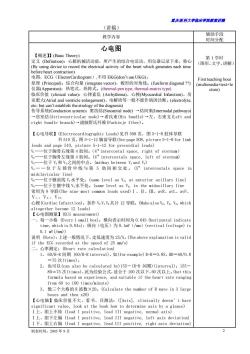
重庆医科大学临床学院教案讲稿 制表时间:2005 年 9 月 2 (讲稿) 教学内容 辅助手段 时间分配 心电图 【概述】】(Basic Theory) 定义 (Definition):心脏机械活动前,所产生的综合电活动,用仪器记录下来,称心 (By using device to record the electrical activity of the heart which generates each time before heart contraction) 电图,ECG(ElectorCardiogram),不用 EKG(don’t use UKG)。 原理 (Principal):综合向量 (integrate vector),梭形的对角线。(fusiform diagonal ??) 仪器(Apparatus):热笔式、热阵式。(thermal-pen type, thermal-matrix type) 临床价值 (clinical value):心律紊乱 (Arrhythmia)、心梗(Myocardial Infarction)、房 室肥大(Atrial and ventricle enlargement)、电解质等一般不能作病因诊断。(electrolyte, etc- but can’t establish the etiology of the diagnosis) 传导系统(Conduction system):窦房结(Sinoatrial node) →结间束(Internodal pathways) →房室结(Atrioventricular node)→希氏束(His bundle)→左、右束支(Left and right bundle branch)→浦倾野氏纤维(Purkinje fiber)。 【心电导联】(Electrocardiographic Leads)见书 508 页,图 5-1-8 肢体导联 书 510 页,图 5-1-12 胸前导联(See page 508, picture 5-1-8 for limb leads and page 510, picture 5-1-12 for precordial leads) V1——位于胸骨右缘第 4 肋间。(4 th intercostal space, right of sternum) V2——位于胸骨左缘第 4 肋间。(4th intercostals space, left of sternum) V3——位于 V2 和 V4 之间的中点。(midway between V2 and V4) V4 — — 位 于 左 锁骨 中 线 与 第 5 肋间 相 交 处 。 (5th intercostals space in midclavicular line) V5——位于腋前线 V4 水平处。(same level as V4, at anterior axillary line) V6——位于左腋中线 V4 水平处。(same level as V4, in the midaxillary line 常用为 9 导联(The nine most common leads used)Ⅰ、Ⅱ、Ⅲ、avR、avL、avF、 Ⅴ1.、Ⅴ3.、Ⅴ5。 心梗(Cardiac Infarction):加作 V2 V4 V5 共计 12 导联。(Make also V2, V4, V5, which altogether become 12 leads) 【心电图测量】(ECG measurement) 一、每一小格 (Every 1 small box):横向表示时间为 0.04S (horizontal indicate time, which is 0.04s);纵向(电压)为 0.1mV(/mm)(vertical (voltage) is 0.1 mV(/mm)) 说明 (Note):上述一般情况下,走低速度为 25/S。(The above explanation is valid if the ECG recorded at the speed of 25 mm/s) 二、心率测定:(Heart rate calculation) 1、 60/R-R 间期 (60/R-R interval),如(for example) R-R=0.8S,HR=60/0.8 =75 次(times)。 2、 也可以(can also be calculated by)155-(R-R 间期)(interval);155- 80=75 次(times),此为经验公式,适合于 100 次以下,60 次以上。(but this formula based on experience, and suitable if the heart rate ranging from 60 to 100 times/minute) 3、 数三个大格的 R 波数×20。(Calculate the number of R wave in 3 large boxes and then x20) 【心电轴】临床价值不大,看书、目测法:([Axis], clinically doesn’t have significant value, look at the book how to determine axis by a glance) Ⅰ上、Ⅲ上不倚 (lead I positive, lead III negative, normal axis) Ⅰ上、Ⅲ下左偏 (lead I positive, lead III negative, left axis deviation) Ⅰ下、Ⅲ上右偏 (lead I negative, lead III positive, right axis deviation) 第 1 学时 (图形、文字、讲解) First teaching hour (multimedia+text+le cture)
重庆医科大半脑床半院载未讲满 第2学时 (图形、文字、讲解 【正常心电图波形及正常值】[Normal wave and value in ECG] Second teaching Pp波:(p wave) 形态(shape):圆钝(smooth rounded)。 text+le 时限(duration):≤0.1 0.25mV R间期(Interval): from the starting point of P wave to starting point of QRS wave is 0.12-0.20s) (can be present or not but ifthere is Qwave,it should be) 时限(duration),<0.04S:振幅<同导联R波的1/4.(the amplitude<1/4 of the amplitude of R wave of the same lead) ST段(segment): j点或Qs波的终点到T波的起点。(J junction or the end of the QRS complex to the beginning of T wave) 压低(depression)0,05sm evat leads)0.1 m m T波(wave) 向多与主波方5。出0 ave):高度应≥1/1OR波】 usually the same as (ta11 ve if 21/10 Rw QT间期(inte a1 随心率而改变(changing accc rding to the heart rate) qTc=qT/R-R0.445。正常值(normal value) I波(wawe). r波之后又一小波(small wave that emerges after T wave) 与T波方向一致,低钾时U波明显增高。(the deflection is the same as T wave in hypokalemia U wave become prominent) 【心房、心室e大】[Left atrial and levt ventricular enlargement] 图诊断是 可靠的,与真实解剖肥大和X线 电压来 (In th 态(shape) rgement): ed P波”,肺心病人常见。(Often termed as"P tient) 左房肥大(left atrial enlargement):形态(shape):P波多呈双峰(double countour P wave), 又称“二尖瓣型P波”,多见于风心病人.(and also termed"P mitrale”,often encountered dise ase patient) 0.12S 心室肥大 largement,P wave become wide and tall) 左心室肥大(eft ventricle dilatation):主要看胸前导联(The most significan changes is in precordial leads) 制表时间:2005年9月
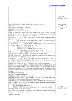
重庆医科大学临床学院教案讲稿 制表时间:2005 年 9 月 3 【正常心电图波形及正常值】[Normal wave and value in ECG] P 波:(P wave) 形态(shape):圆钝(smooth rounded)。 时限 (duration):≦0.11S 振幅 (amplitude):0.2~0.25 mV P-R 间期 (Interval):从 P 波开始,到 QRS 波开始的时间为 0.12~0.20S。(The time from the starting point of P wave to starting point of QRS wave is 0.12-0.20s) QRS 波群 (complex): 时限(duration)<0.12S Q 波(wave): 可有、可无。如有 Q 波应是:(can be present or not, but if there is Q wave, it should be) 时限 (duration)<0.04S;振幅<同导联R波的1/4。(the amplitude<1/4 of the amplitude of R wave of the same lead) ST 段 (segment): j 点或 QRS 波的终点到 T 波的起点。(J junction or the end of the QRS complex to the beginning of T wave) 压低(depression)≯0.05S mV; 抬高(elevation):肢体导联(limb leads)≯0.1 mV Ⅴ1.、Ⅴ2≯0.3SmV、Ⅴ3≯0.5 mV、Ⅴ5-6≯0.1 mV T 波 (wave): 方向多与主波方向同 (the defelection usually the same as the QRS wave);高度应≥1/10R 波。(tall wave if ≥1/10 R wave) QT 间期 (interval): 随心率而改变 (changing according to the heart rate)。 QTC=QT/R-R 0.44S。正常值 (normal value) U 波(wave): T 波之后又一小波 (small wave that emerges after T wave) , 与 T 波方向一致,低钾时 U 波明显增高。(the deflection is the same as T wave, in hypokalemia U wave become prominent) 【心房、心室肥大】[Left atrial and levt ventricular enlargement] 说明 (Note):心房、心室肥大的 心电图诊断是不可靠的,与真实解剖肥大和 X 线 等比较,敏感性和特异性都差,临床多用高电压来表达。(In the case of left atrial and left ventricle enlargement, ECG is unreliable. Compared to the actual size and X ray, ECG has lower sensitivity and specificity. In clinical practice, usually we expressed it as high voltage. 心房肥大(atrial enlargement) : 右房肥大 (right atrial enlargement): 形态(shape):高尖。(tall peaked and pointed ) 振幅(amplitude)≥0.125 mV,又称为“肺型 P 波”,肺心病人常见。(Often termed as “P Pulmonale”, and often encountered in cor pulmonale’ patient) 左房肥大(left atrial enlargement):形态(shape):P 波多呈双峰(double countour P wave), 又称“二尖瓣型 P 波”,多见于风心病人。(and also termed “P mitrale”, often encountered in rheumatic heart disease’ patient) 增宽 (prolonged duration)≥0.12S。 双房肥大,增宽、增高(biatrial enlargement, P wave become wide and tall) 心室肥大 (Ventricular Dilatation) 左心室肥大(left ventricle dilatation):主要看胸前导联 (The most significant changes is in precordial leads) 第 2 学时 (图形、文字、讲解) Second teaching hour (multimedia+text+le cture) 1 学时 (图形、文字、讲解)

露庆医科大学临床半蕊藏案讲满 Rs≥2.5mV:Rs.S=4mV:awF≥2mV: 如有ST改变,称为左室肥大伴劳损(if accompanied by ST change, hing hour diagnosed as left ventricular enlargement accompanied by injury),临床上如没有ST改变,就直接写左室高电压。(ifclinically cture) doesn't have ST change,diagnosed as left ventricular high ricle dilatation):VRs≥】 of both loft icle nd ight 【心肌缺血】(Cardiac ischemia) T波对称性倒置,又称“冠状T”(inverted symmetrical T wave,.called as “coronary T wave ST改变(ST change),为非特异性(unspecific)。冠心病、心肌炎等都可以有此 表现,但如果是区域性改变,结合临床,多考虑为冠心病。(Both coronary heart disease nd myocardis can c ST change,but coronary heart disease should be considered 临床意义(clinical significance):心肌梗塞多见,临床情况严重,早诊断早治疗可获 得较好的效果,时间就是生命。(myocardial infarction is commonly found in clinica practice,usually have severe presen 华方快的 心电图演变过程:ECGvolution proces) ·超急期:(hyperacute phase) 主要是T波改变,变得高尖或R波的改变,ST段抬高或正常,呈区域 性改变。(The most prominent change is that T wave become tal and pointed or R wave change,with elevated or normal ST segment, ·急性期 心电图可以有典型的改变,或者说经典改变。E can have typical hange) ※出现病理性Q波()≥0.04S(found out pathologic oa ≥0.O4S,深度≥1/4R波或呈qS型。(amplitude≥1/4 R or found ※ST弓背抬高,呈单向曲线。(ST segments evolve from concav, to a convex upward pattern to a unidrecional curve) ·亚急性期:(Subacute phase) ※数天、数周,少数可达数月.(a few days or weeks,rarely reach a fev months) radually return to 1 inverte d als ur to normal) ※坏死型Q波继续存在。(but pathologic Q wave remain)) 制表时间:2005年9月
重庆医科大学临床学院教案讲稿 制表时间:2005 年 9 月 4 RV5 ≥2.5 mV;RV5 =SV1=4mV;avF ≥2mV; 如有 ST 改变,称为左室肥大伴劳损(if accompanied by ST change, diagnosed as left ventricular enlargement accompanied by injury),临床上如没有 ST 改变,就直接写左室高电压。(if clinically doesn’t have ST change, diagnosed as left ventricular high voltage) 右室肥大(right ventricle dilatation):Ⅴ1.R/S≥1 双室肥大 (Biventricular dilatation): 既有左心室肥 大的心电图 表现,也有 右心室心 电图表现。(Have both the characteristics of both left ventricle and right ventricle dilatation) 【心肌缺血】(Cardiac ischemia) T 波对称性倒置,又称“冠状 T”(inverted symmetrical T wave, called as “coronary T wave” ST 改变 (ST change),为非特异性 (unspecific)。冠心病、心肌炎等都可以有此 表现,但如果是区域性改变,结合临床,多考虑为冠心病。(Both coronary heart disease and myocarditis can cause ST change, but coronary heart disease should be considered firstly for the regional ST change) 【心肌梗塞】[Myocardial Infarction] 临床意义(clinical significance):心肌梗塞多见,临床情况严重,早诊断早治疗可获 得较好的效果,时间就是生命。(myocardial infarction is commonly found in clinical practice, usually have severe presentation, early diagnosis and early treatment can improve prognosis, time is life) ECG 是最简单、最方便、最快捷的诊断,在心梗诊断 中,占有十分重要的地位。(ECG is the easiest, the most efficient and the quickest tools for diagnosis. It has important role in the diagnosis of myocardial infarction) 心电图演变过程:(ECG evolution process) • 超急期:(hyperacute phase) 主要是 T 波改变,变得高尖或 R 波的改变,ST 段抬高或正常,呈区域 性改变。(The most prominent change is that T wave become tall and pointed or R wave change, with elevated or normal ST segment, regionally.) • 急性期:(Acute phase) 心电图可以有典型的改变,或者说经典改变。(ECG can have typical change) ※ 出现病理性 Q 波(宽)≥0.04S (found out pathologic Q wave ≥0.04s),深度≥1/4R 波或呈 QS 型。(amplitude ≥1/4R or found out QS wave) ※ ST 弓背抬高,呈单向曲线。(ST segments evolve from concave to a convex upward pattern to a unidrecional curve) • 亚急性期:(Subacute phase) ※ 数天、数周,少数可达数月。( a few days or weeks, rarely reach a few months) ※ 抬高的ST段逐渐到等电线,倒置T波亦逐渐恢复正常。(ST segment gradually return to isoelectric line, inverted T wave also gradually return to normal) ※ 坏死型 Q 波继续存在。(but pathologic Q wave remain) one teaching hour (multimedia+text+le cture)
重庆医科大半脑床半院载未讲满 ·陈旧型心梗:(Old Myocardial Infarction) ※3-6月,病理性Q波,ST波恢复正常或继续T波倒置。(3-6 months pathologic Q wave,ST segment return to normal ※病理性Q波(pathologic Q wave) ※ST-T恢复正常或持续倒置。(ST-T return to normal or 心肌梗塞的定位:(The location of myocardial infarction) 说明:(Note) 3学时 1、异常Q波不一定是心肌梗塞Q波(Abnormal Q wave is not necessarily (图形、文字、讲解 myocardial infarction)。脑血管意外、肺气肿等者 2、 心肌梗塞从ECG分类可分为(From ECG,myocardial infarction can be cture) ·非Q波型心肌柜塞 ·正常EOG型心肌梗塞( infarction) infarction) 律失常(Cardiac arrhythmia) 意义(Clinical significance)):心律失常是ECG唯一的诊断,也是ECG的最大价值 也是ECG最复杂的部分(Cardiac arrhythmia is an ECG diagnosis,also 【概述】(General explanation) 引用书529页的表格 【正常心电图】(Nom al ECG) 的(PQRS-Twa segmen fthe ry ular) 6、QRS波的宽度与高度正, and amplitude of QRS complex is normal) 7、心率60-100次/分(Heart rate60-100 times/minute) 8、无病理性O被There is no pathologic O wave) 【异常窦性心电图】(Abnormal sinus ECG) 1、窦性窦性心动过速>00次/min(Sinus Tachycardia)100 times/minute) 2、窦性窦性心动过缓<60次/min(Sinus Bradycardia<60 times/minute) 制表时间:2005年9月
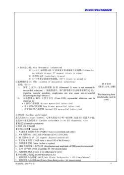
重庆医科大学临床学院教案讲稿 制表时间:2005 年 9 月 5 • 陈旧型心梗:(Old Myocardial Infarction) ※ 3-6 月,病理性 Q 波,ST 波恢复正常或继续 T 波倒置。(3-6 months, pathologic Q wave, ST segment return to normal ※ 病理性 Q 波 (pathologic Q wave) ※ ST-T 恢复正常或持续倒置。(ST-T return to normal or 心肌梗塞的定位:(The location of myocardial infarction) 说明:(Note) 1、 异常 Q 波不一定是心肌梗塞 Q 波 (Abnormal Q wave is not necessarily myocardial infarction) 。脑血管意外、肺气肿等都可以出现非梗塞性 Q 波。 (Cerebral vascular accidents, emphysema can also cause non-myocardial infarction pathologic Q wave.) 2、 心肌梗塞从 ECG 分类可分为 (From ECG, myocardial infarction can be classified as) • Q 波型心肌梗塞 (Q wave myocardial infarction) • 非 Q 波型心肌梗塞 (non Q wave myocardial infarction) • 正常 ECG 型心肌梗塞 (normal ECG myocardial infarction) 心律失常 (Cardiac arrhythmia) 意义(Clinical significance):心律失常是 ECG 唯一的诊断,也是 ECG 的最大价值, 也是 ECG 最复杂的部分 (Cardiac arrhythmia is an ECG diagnosis, also 【概述】(General explanation) 引用书 529 页的表格 【正常心电图】(Normal ECG) 1、P-QRS-T 波是相关的 (P-QRS-T wave is correlated each other) 2、P-R 间期≥0.12S100 次/min (Sinus Tachycardia > 100 times/minute) 2、窦性窦性心动过缓<60 次/min (Sinus Bradycardia < 60 times/minute) 第 3 学时 (图形、文字、讲解) Third teaching hour (multimedia+text+le cture)
露庆医科大学临床半院载案讲满 3、窦性心率不齐R-R间期之差>0.l2S(Sinus arrhythmia,the differences of R-R interval>0.12 s) 4、窦性暂停或停搏,P搏消失,出现结性或室性逸搏性心律。(Sinus pause or sinus 说明:(note) 配对间期(联律间期):早搏与其前窦性QRS波的时距,(Coupling Interval:The interval 个窦性QRS搏的时距(Compensatory pause:The interval 供完m配对代隙两不常的R间期((Fully com ensatory pause >R-R interval of the two consecutive normal sinus beat) 代偿不完全:配对间期+代偿间隙0.12S) ·代偿完全(fully compensatory pause) ·未打乱窦性节律 二、房性过早搏动(Atrial premature complex) ·提前出现的 -QRS- 波群,P'波与窦性P有差异(There is P-QRS-T wave complex,the of the wave is di ferent from normal P wave) interval≥0.12 、结性过早短动unctional。 ·提前出现的QRS波群,QRS波为室上性,其P波有三种情况(There is QRS wav: complex.the ors is supraventricular.There are 3 possibilities of p wave ①QRS波之前②在之后③无P波(L.Before QRS wave2.After QRS wave 3 There is no p wave) 制表时间.2005年9月
重庆医科大学临床学院教案讲稿 制表时间:2005 年 9 月 6 3、窦性心率不齐 R-R 间期之差>0.12S (Sinus arrhythmia, the differences of R-R interval>0.12 s) 4、窦性暂停或停搏,P 搏消失,出现结性或室性逸搏性心律。(Sinus pause or sinus arrest, P wave disappear, and there is junctional or ventricle escape rhythm) 【过早搏动或期前收缩】 说明:(note) 配对间期(联律间期):早搏与其前窦性QRS波的时距,(Coupling Interval : The interval between premature beat and the previous sinus QRS wave) 代偿间隙:早搏后与第一个窦性 QRS 搏的时距 (Compensatory pause: The interval between premature beat to the first next sinus QRS wave) 代偿完全:配对间期+代偿间隙≥两个正常的 R-R 间期 (Fully compensatory pause : coupling interval + compensatory pause ≥ R-R interval of the two consecutive normal sinus beat) 代偿不完全:配对间期+代偿间隙0.12S) • 代偿完全 (fully compensatory pause) • 未打乱窦性节律 二、房性过早搏动 (Atrial premature complex) • 提前出现的 P ′-QRS-T 波群,P ′波与窦性 P 有差异 (There is P-QRS-T wave complex, the shape of the P wave is different from normal P wave) • P ′-R 间期≥0.12S (P-R interval≥0.12S) • 代偿不完全 (There is no fully compensatory pause) • QRS 为室上性,与窦性 QRS 相同 (The QRS wave is supraventricular and have similar shape as the sinus QRS wave) 三、结性过早搏动 (Junctional premature complex) • 提前出现的 QRS 波群,QRS 波为室上性,其 P 波有三种情况 (There is QRS wave complex, the QRS is supraventricular, There are 3 possibilities of P wave ① QRS 波之前 ②在之后 ③无 P 波 (1. Before QRS wave 2. After QRS wave 3 There is no P wave) 第 4 学时 (图形、文字、讲解) Fourth teaching hour (multimedia+text+le cture)
重庆医科大学脑床半院载未讲满 ·如果有P波,无论在之前或之后均为倒置,P-R或R-R均<0.12S(If there is P wave,either before or after QRS wave,the distance of P-R or R-P <O.12S ·代偿完全(Fully compensatory pause)) 【心动过速】(Tachycardia) 过速包括房、结、室性心动过速,因发热或出血等引起的心动过 太太t进花遇内:betru,veiricle ty回 发性密上性,心,动过速P3 Supr ventricular Tachy 阵发性心动过速现应包括房性和结性(Paroxysmal Supraventricular Tachycardia on sists of either atrial or junctional)),但心动过速发生后,P'波落在前一个Qs波的T 波上难以区别房或结性(but if when the tachycardia emerges,.and there is P wave before QRS but inside the T wave,then it is difficult to differentiate between atrial and junctional),同时处理都基本一致,因此统称为室上性心动 (but still the mans thus usually Just termed a 1学时 (图形、文字、讲解 折返学说:结内折返7O%(可用洋地黄)(reentry:AV node reentry70%(Ca se digitalis) one teaching hour 旁道折返20%(预激,不能用洋地黄)(additional pathway reentry20% (multimedia+text+le (preexcitation,can't use digitalis) cture) ECG特点:(ECG characteristics) 1、突发、突止。 (Appear suddenly and disappear suddenly) 2、节律规则,频率多在160~250次。(Regular rhythm,.rate increase, 波为o160250ties (supraventricular QRS) 临床是较为餐的C nore fatal) 连续出现宽大畸型的ORS波群。 (The e are cor secutive wide and bizarre QRS a、节律提则,频率在10-20次/分((Re证rateyrond 子、成不可LP涉但p与ORs波无关(Can have or don't have Pwave,.bu 三、室性扑动(Ventricle Flutter:临床上更紧急,ECG特点,ORS波形态消失,必 连续的正弦曲线。200-250次/分。(Clinically even more fatal,.ECG characteristic there is no QRS wave anymore 续的宽 最为紧急 可引起AS综合症,ECG特点:道 卜规则 、AA S波群20 -250次/分.(the most fatal 五、扭转型室速Torsade de Pointes:)室速以基线为轴,ORS波发生上、下方向有 定规律的改变.临床需紧急处理,易发生房颜。(①ype of ventricular tach ardia in which isoelectric line serves as axis and there is gradual rhythmic char nge in the pitd the e be ave 六碳:角床上多是的6律表乱之一6m时血e EC 1、 E350-600次/分(Ther ave and replaced by a wave tha ent shane and size 3 ORS波为室上性(Su 4、临床上多有脉搏短促现象。(Clinically the pulse is short and fast) 制表时间:2005年9月
重庆医科大学临床学院教案讲稿 制表时间:2005 年 9 月 7 • 如果有 P 波,无论在之前或之后均为倒置,P ′-R 或 R ′ -R 均<0.12S(If there is P wave, either before or after QRS wave, the distance of P-R or R-P <0.12S • 代偿完全 (Fully compensatory pause) 【心动过速】 (Tachycardia) 说明 (note):心动过速包括房、结、室性心动过速,因发热或出血等引起的心动过 速不在此讲范围内。(Tachycardia can be either atrial, junctional, or ventricle tachycardia, tachycardia because of fever, or bleeding is not included in this group) 一、阵发性室上性心动过速 (Paroxysmal Supraventricular Tachycardia) 阵发性心动过速现应包括房性和结性 (Paroxysmal Supraventricular Tachycardia consists of either atrial or junctional),但心动过速发生后,P ′波落在前一个 QRS 波的 T 波上难以区别房或结性 (but if when the tachycardia emerges, and there is P wave before QRS but inside the T wave, then it is difficult to differentiate between atrial and junctional) ,同时处理都基本一致,因此统称为室上性心动 过速。(but still the management is similar, thus usually just termed as supraventricular tachycardia) 机制:(mechanisms) 独立兴奋点<50% (automaticity<50%) 折返学说:结内折返 70%(可用洋地黄)(reentry: AV node reentry 70% (Can use digitalis) 旁道折返 20%(预激,不能用洋地黄)(additional pathway reentry 20% (preexcitation, can’t use digitalis) ECG 特点:(ECG characteristics) 1、 突发、突止。(Appear suddenly and disappear suddenly) 2、 节律规则,频率多在 160~250 次。(Regular rhythm, rate increase, usually around 160-250 times) 3、 QR 波为室上性。(supraventricular QRS) 二、室性心动过速 (Ventricular Tachycardia) 临床上是较为紧急的 (Clinically more fatal) 1、连续出现宽大畸型的 QRS 波群。(There are consecutive wide and bizarre QRS wave) 2、 节律规则,频率在 140-200 次/分 (Regular rhythm, rate usually around 140-200 times/minute) 3、可见或不可见 P 波,但 P 与 QRS 波无关 (Can have or don’t have P wave, but there is no relation between P wave and QRS wave) 三、室性扑动 (Ventricle Flutter):临床上更紧急,ECG 特点,QRS 波形态消失,必 连续的正弦曲线。200-250 次/分。(Clinically even more fatal, ECG characteristic, there is no QRS wave anymore .) 四、心室颤动(Ventricle Fibrillation):最为紧急,可引起 A-S 综合症,ECG 特点:连 续的宽大畸型、大小不等节律不规则的 QRS 波群 200-250 次/分。(the most fatal, can cause Adam Stroke/A-S syndrome, ECG characteristic: .) 五、扭转型室速(Torsade de Pointes):室速以基线为轴,QRS 波发生上、下方向有一 定规律的改变。临床需紧急处理,易发生房颤。(Type of ventricular tachycardia, in which isoelectric line serves as axis and there is gradual rhythmic change in the amplitude (changing between up and down) and twisting of the QRS wave. Clinically, it needs to be treated quickly, since it frequently degenerate become ventricular fibrillation) 六、房颤 (Atrial Fibrillation):临床上多见的心律紊乱之一(It is one of the most common arrhythmia encountered in clinical practice),ECG: 1、 P 波消失,代之以大小不等,形态各异的“f”波,频率在 350-600 次/分(There is no P wave and replaced by a wave that have different shape and size termed as “f” wave. The rate is around 350-600 times/minute) 2、 QRS 波之间绝对不规则 (The interval between QRS wave is not regular) 3、 QRS 波为室上性 (Supraventicular QRS wave) 4、 临床上多有脉搏短促现象。(Clinically the pulse is short and fast) 1 学时 (图形、文字、讲解) one teaching hour (multimedia+text+le cture)
露庆医科大学临床半院载案讲满 【传导阻滞】(Conduction block) 传导系统任何部位都可以发生阻滞,临床上以房室结和心室内阳最为常见 tion hlock in the he moxceommomconductionbiockisalrnoelriulrblbckadintrarcnlicdarbloCk 房室传 0G特点 -R间期≥0.21S characteristic:P-R interval>0.21S 2、 21 文氏型(Wenkebach type):P-R间期逐渐延长, 直至一个RS油 后P-R间期趋于缩短,再有 一个逐海延,周而复始。(P-R interva】 gradually increases until suddenly there is one drop of QRS wave Then the p-r interval become short again and gradually increases and there is another drop.It repeated overtime) 2)莫氏型(莫氏Ⅱ型)(Mobitz type I川):PR间期固定,出现不固定的QRS波 脱漏现象。 (P-R interval remain the same but suddenly there is one ve 阻滞,P 完全无关, 波多 波 态取决 再右心 如再 告就是室上性 性心律 见 (AI relation be of the Ops da of the ck.fo if the hlock in the al node then it shane like su scape rhythm:if the block in right ventricle then the QRS become wide and hizarre) 三、束支阻滞(Bundle branch block) 左束支阻滞:(Left bundle branch block) I、完全性左束支阻滞(Complete left bundle branch block) QRS波>0.12S(QR e>0.125) M"型。(R wave of the lead V have a notch or have金 导联的R波有切迹 左束支 complete left bundle branch block):QRS.12 R波有切迹或呈 ndle or d V,have a notchor have 导联 shape 1、完全性右束支阻滞(Co tight bundle branch block): QR5波>0.12S(QR8 e0.l2S):,导联的R波呈r'sR型。(R wave of the lead V,have the shap of r'sR') 2、不完全性右束支阻滞Incomplete right bundle branch block):QRS0.12S(QRS wave>0.12S): 但QRs波形既不像左束支也不像右束支阻滞图形。(but QRS wave didn't胎tch iter 1、PR间期PRin 31<019 2、R波起始部分有挫折(Delta波,△波)(there is slurred upstroke at the beginning of the QRS wave (delta wave)) QRS波≥0.12 ave≥0.12S 又称P.R间期缩短综合征。ames known as 制表时间:2005年9月 8
重庆医科大学临床学院教案讲稿 制表时间:2005 年 9 月 8 【传导阻滞】(Conduction block) 传导系统任何部位都可以发生阻滞,临床上以房室结和心室内阻滞最为常见。 (Conduction block can occurred at any conduction system in the heart. Clinically, the most common conduction block is atrioventricular block and intraventricular block) 一、房室传导阻滞 (Atrioventricular Block) 1、Ⅰ 。-AVB (Ⅰ 。房室传导阻滞)(I° Atrioventricular Block) ECG 特点:P-R 间期≥0. 21S (ECG characteristic: P-R interval≥0. 21S 2、Ⅱ 。-AVB 1) 文氏型(Wenkebach type):P-R 间期逐渐延长,直至脱漏一个 QRS 波, 后 P-R 间期趋于缩短,再有一个逐渐延,周而复始。(P-R interval gradually increases, until suddenly there is one drop of QRS wave. Then the P-R interval become short again and gradually increases and there is another drop. It repeated overtime) 2) 莫氏型(莫氏Ⅱ型)(Mobitz type II):P-R 间期固定,出现不固定的 QRS 波 脱漏现象。(P-R interval remain the same but suddenly there is one drop of QRS wave) 3、Ⅲ。-AVB:又称为完全性传导阻滞,P 波与 QRS 完全无关,往往 P 波多于 QRS 波, 其 QRS 形态取决于阻滞起博点位置,如再房室结就是室上性,结性逸搏性心律;如 再右心室就是宽大畸型的 QRS 波。(Also termed as complete AV block. There is no relation between P and QRS wave. Usually it has more P wave than QRS wave,the shape of the QRS depends on the location of the conduction block, for example if the block in the AV node then it shape like supraventricular, junctional escape rhythm; if the block in right ventricle then the QRS become wide and bizarre) 三、束支阻滞 (Bundle branch block) (一)左束支阻滞:(Left bundle branch block) 1、完全性左束支阻滞(Complete left bundle branch block): QRS 波>0.12S(QRS wave>0.12S);V5 导联的 R 波有切迹或呈“M”型。(R wave of the lead V5 have a notch or have “M” shape) 2、不完全性左束支阻滞(Incomplete left bundle branch block):QRS0.12S(QRS wave>0.12S);V1 导联的 R 波呈 r'sR'型。(R wave of the lead V1 have the shape of r'sR') 2、不完全性右束支阻滞(Incomplete right bundle branch block):QRS0.12S (QRS wave>0.12S); 但 QRS 波形既不像左束支也不像右束支阻滞图形。(but QRS wave didn’t match for the criteria of either complete left bundle branch block or complete right bundle branch block) 三、预激综合症 (Preexcitation Syndrome) 因旁道原因。传导未经房室结或部分房室结所致。 (一) 典型预激(WPW)(Kent preexcitation) 1、P-R 间期 (P-R interval)<0.12S 2、R 波起始部分有挫折(Delta 波,△波)(there is slurred upstroke at the beginning of the QRS wave (delta wave)) 3、 QRS 波≥0.12S;(QRS wave≥0.12S) (二) J ame’s 预激,又称 P-R 间期缩短综合征。(James preexcitation or also known as short P-R interval syndrome) 1、P-R 间期 (P-R interval)<0.12S
置庆医科大半脑床半院未讲满 2、QRS波正常,无Delta波。(QRS wave normal,there is no delta wave) (三)Mahain预激(Mahain preexcitation) I、P-R间期正常,(P-R interval normal) (QRS wave>0.12S) 3、QRs波起始部可见Delta波。(There is delta wave at the beginning of QRS) 其它常用心电图学检查(The other ECG examination that frequently used) (AECG)又名 原理(Pr () 回放(Analysis):不可能用眼去阅读,而采用计算机技术,把数字图形化,设计临床 需要的软件,例如:室性过早搏动,ST改变等,通过快速回放,就将所周 的资料报告出来。(Can't be analyzed just by using eye.It uses computerized em that b shape.And then i ventricular compexorTseement.Then the resut will automaically come out) 临床价值:(Clinical 大的月 用途。(Can be used fo short periode of rhvthm dirint use of AECG) the present of cardiac ischemia) 3、晕厥、胸闷等短暂出现的症状临床的判断。(To diagnose syncope,.chest discomfort and other short periode clinical symptoms) 原理动试 耗氧量增加(心律加快)一冠状血流正常一ST无改变 当冠状血管病变狭窄时冠流不能随耗氧量(心率)增加而增加时ST下移( 即缺血性改变。 blood flov e m is no ch (H there is sten of the coronary bloo oxygen ange 方法.Method) (一)活动平板(treadmill)。 l、原理(Principal):相当一个跑步机,有坡度加速度,随着运动量加量坡度越来越 大,速度越来越快速,比较经典的就是美国人Buce方案(布朗氏方案)(Th most frequent is by using Bruce method) 2 结果判定(():ST水平或重型压低01mv为阳(性6 1、原理P 的是压级量心率,常采用公式155 一年龄 (.(Work load will gradually increases until reach target heart rate 155-age) 3、 2、结果判断(Result interpretation:)ST水平或下重型ST压低0.lmv为阳性。(ST depression that reach.1 mv considered as positive) 三)智能式冠心病检查仪CASE-Computerized Analysing System for Exercise) veloped in the past 2 years) variable and ST dependent variable). 制表时间:2005年9月
重庆医科大学临床学院教案讲稿 制表时间:2005 年 9 月 9 2、QRS 波正常,无 Delta 波。(QRS wave normal, there is no delta wave) (三) Mahain 预激 (Mahain preexcitation) 1、P-R 间期正常,(P-R interval normal) 2、QRS 波>0.12S (QRS wave>0.12S) 3、QRS 波起始部可见 Delta 波。(There is delta wave at the beginning of QRS) 其它常用心电图学检查 (The other ECG examination that frequently used) 一、动态心电图 ambulatory electorcardiography(AECG)又名 Aoleer (Holter) 原理(Principal):记录系统,将24小时的心电信号数字化,记录在磁带上。(Continuously recording the electrical activity of the heart for 24 hours in a cassette) 回放 (Analysis):不可能用眼去阅读,而采用计算机技术,把数字图形化,设计临床 需要的 软件,例如:室性过早搏动,ST 改变等,通过快速回放,就将所需 的资料报告出来。(Can’t be analyzed just by using eye. It uses computerized system that being adjusted and change the data become a shape. And then it adjusted to specific abnormalities that want to be analyzed such as premature ventricular complex or ST segement. Then the result will automatically come out) 临床价值:(Clinical value) 1、各种心律紊乱,有无长间隙,这是 AECG 最大的用途。(Can be used fo short periode of rhythm disorder. It is the most important use of AECG) 2、有无 性缺血(S、M、I 现象-Silence Myocardial ischemic (To analyze the present of cardiac ischemia) 3、晕厥、胸闷等短暂出现的症状临床的判断。(To diagnose syncope, chest discomfort and other short periode clinical symptoms) 二、运动试验 (Exercise Test) 原理(Principal):运动→心肌耗氧量增加(心律加快)-冠状血流正常-ST 无改变。 当冠状血管病变狭窄时冠流不能随耗氧量(心率)增加而增加时 ST 下移(压 低),即缺血性改变。(Exercise makes increase oxygen consumption (Heart rate increase), If coronary blood flow normal, there is no change in ST segment, but if there is stenosis of the coronary artery, there is decrease of the coronary blood flow and make increase of the oxygen consumption and it will make increase of the heart rate and ST segment depression, change of ischemic heart disease 方法:(Method) (一)活动平板(treadmill)。 1、 原理(Principal):相当一个跑步机,有坡度加速度,随着运动量加量坡度越来越 大,速度越来越快速,比较经典的就是美国人 Bruce 方案(布朗氏方案)(The principal is like a machine where pople can run and can increase in speed. The patient will be given some workload that will increase in time and also become faster, or the most frequent is by using Bruce method) 2、 结果判定(Result interpretation):ST 水平或重型压低 0.1mv 为阳性(ST depression that reach 0.1 mv considered as positive) (二)踏车试验(bicycle) 1、原理(Principal):有一功量计,逐渐增加运动量,直至达靶心率,目前使用最多 的是压级量心率,常采用公式 155-年龄。(.) (Work load will gradually increases until reach target heart rate 155-age) 3、 2、结果判断(Result interpretation):ST 水平或下重型 ST 压低 0.1mv 为阳性。(ST depression that reach 0.1 mv considered as positive) (三)智能式冠心病检查仪 CASE-Computerized Analysing System for Exercise) 1、 原理(Principal):它是运动试验基础上(by using exercise test),最近 20 年度发展起 来的一种新的冠心病检查方法(It is a new method for coronary heart disease examination which has been developed in the past 20 years) ,是以心率为自变量, ST 为应变量的回归直线 (It is a linear regression that use heart rate as independent variable and ST as dependent variable),又称 ST/HR 斜率法 (Also called ST/HR
露庆医科大学临床半院载案讲满 slope method,或心率矫正ST法(or Heart rate modification ST method)。 I)ST段压低程度(Degree of ST depression)=心肌缺血严重程度.(Ischemic degree of 分食大服R I、结果判断(Interpretation):ST/HR.Slopet(STHR斜率)uv/bpm为单位(As unit)). 8 生别 y93 假阳性合假阴性,书上554页的评论。( and biycl)veiher falspoiand 制表时间:2005年9月 10
重庆医科大学临床学院教案讲稿 制表时间:2005 年 9 月 10 slope method),或心率矫正 ST 法 (or Heart rate modification ST method)。 1)ST 段压低程度 (Degree of ST depression)=心肌缺血严重程度.(Ischemic degree of myocardium)×运动负荷大小(Work load)(心率(heart rate)-HR)。 2)心肌缺血程度(Ischemic degree of myocardium)=ST/HR. 1、结果判断(Interpretation):ST/HR.Slope(ST/HR 斜率) uv/bpm 为单位(As unit)。 2、 优点(Advantage): (1) 敏 感 性 和 特 异性 较 高 (Higher sensitivity and specificity) , 敏 感 性 (Sensitivity)93~96%;特异性(specificity)93~97% (2) 不受药物、性别影响。(Not influenced by medication and gender) (3) 传统的运动试验(活动平板或踏车)的敏感性合特异性较低,有较多的 假阳性合假阴性,书上 554 页的评论。(the previous exercise test(treadmill and bicycle) have higher false positive and false negative)
按次数下载不扣除下载券;
注册用户24小时内重复下载只扣除一次;
顺序:VIP每日次数-->可用次数-->下载券;
- 重庆医科大学:《诊断学》课程教学资源(授课教案)06 腹泻.doc
- 重庆医科大学:《诊断学》课程教学资源(授课教案)05 腹痛.doc
- 重庆医科大学:《诊断学》课程教学资源(授课教案)07 黄疸.doc
- 重庆医科大学:《诊断学》课程教学资源(授课教案)02 水肿.doc
- 重庆医科大学:《诊断学》课程教学资源(授课教案)01 绪论与问诊.doc
- 重庆医科大学:《诊断学》课程教学资源(授课教案)08 腹部检查.doc
- 重庆医科大学:《诊断学》课程教学资源(授课教案)09 病历与诊断.doc
- 重庆医科大学:《诊断学》课程教学资源(授课教案)03 呕血.doc
- 重庆医科大学:《诊断学》课程教学资源(授课教案)04 便血.doc
- 重庆医科大学:《实验诊断学》课程教学资源(PPT课件)第八讲 脑脊液常规及生殖系统检查.ppt
- 重庆医科大学:《实验诊断学》课程教学资源(PPT课件)第七讲 大便常规、免疫学检查及心肌标志物检查.ppt
- 重庆医科大学:《实验诊断学》课程教学资源(PPT课件)第六讲 肾功能检查(主讲:唐敏).ppt
- 重庆医科大学:《实验诊断学》课程教学资源(PPT课件)第四讲 血栓与出血检查(主讲:胥文春).ppt
- 重庆医科大学:《实验诊断学》课程教学资源(PPT课件)第三讲 骨髓细胞学检查、血型与输血.ppt
- 重庆医科大学:《实验诊断学》课程教学资源(PPT课件)第一讲 总论及血液一般检查(上).ppt
- 上海科学技术出版社:《实验诊断学——彩色图谱》书籍PDF电子版(主编:张丽霞、陈金宝).pdf
- 重庆医科大学:《实验诊断学》课程复习题集(无答案).doc
- 《实验诊断学》课程实验指导书 laboatoty diagnosis(共九章,含练习及答案).doc
- 重庆医科大学:《实验诊断学》课程教学资源(教案)第8讲 CSF生殖系统.doc
- 重庆医科大学:《实验诊断学》课程教学资源(教案)第7讲 大便 免疫 心肌.doc
- 重庆医科大学:《诊断学》课程教学资源(授课教案)11 心脏检查.doc
- 重庆医科大学:《诊断学》课程教学资源(授课教案)20 呼吸系统症状和体征.doc
- 重庆医科大学:《诊断学》课程教学资源(授课教案)13 循环系统主要症状与体征.doc
- 重庆医科大学:《诊断学》课程教学资源(授课教案)15 超声心动图检查.doc
- 重庆医科大学:《诊断学》课程教学资源(授课教案)12 血管检查.doc
- 重庆医科大学:《诊断学》课程教学资源(授课教案)10 发热.doc
- 重庆医科大学:《诊断学》课程教学资源(授课教案)19 胸部检查.doc
- 重庆医科大学:《诊断学》课程教学资源(授课教案)18 呼吸困难.doc
- 重庆医科大学:《诊断学》课程教学资源(授课教案)16 咳嗽、咳痰.doc
- 重庆医科大学:《诊断学》课程教学资源(授课教案)17 咯血.doc
- 医学临床本科《MRI诊断学》课程教学大纲 Magnetic Resonance Imaging Diagnosis.doc
- 医学临床本科《影像诊断学》课程教学大纲 medical imaging(包含MRI部分).doc
- 医学影像本科《MRI诊断学》课程教学大纲(Magnetic Resonance Imaging Diagnosis).doc
- 石河子大学:《MRI诊断学》课程教学资源(讲稿,共五章).doc
- 石河子大学:《MRI诊断学》课程教学课件(PPT讲稿)第一章 MRI总论.ppt
- 石河子大学:《MRI诊断学》课程教学课件(PPT讲稿)第三章 呼吸系统 3.1 3.2 正常及异常.ppt
- 石河子大学:《MRI诊断学》课程教学课件(PPT讲稿)第三章 呼吸系统 3.3 支气管扩张及肺部炎症.ppt
- 石河子大学:《MRI诊断学》课程教学课件(PPT讲稿)第三章 呼吸系统 3.4 肺结核.ppt
- 石河子大学:《MRI诊断学》课程教学课件(PPT讲稿)第三章 呼吸系统 3.5 肺肿瘤.ppt
- 石河子大学:《MRI诊断学》课程教学课件(PPT讲稿)第三章 呼吸系统 3.6 纵膈疾病.ppt
