重庆医科大学:《诊断学》课程教学资源(授课教案)08 腹部检查
置庆医科大学脑床半院藏来讲满 重庆医科大学临床学院教案及讲稿 课程名称诊断学 年级2005 授课专业临床医学 教师古赛 职称副教授 授课方式 大课 学时4 题目章节腹部检查 教材名称诊断学(双语》 作者 吕卓人雷寒 出版社科学出版社 版次第一版 1 To understand the corresponding relationship between abdominal subregions and abdominal splanchna 教学目的要求 2. To be familiar with the contents of abdominal inspection and auscultation 3. To master the contentsand methods of abdominal palpation and percussion To master the palpation and clinical significance of normal and abnormal liver and spleen 5. To master the points of palpation of abdominal masses 教学难点 the palpation and clinical significance of normal and abnormal liver and spleen The clinical significance of abdominal distension,abdominal veins,peristalsis,shifting dullness and change of bowel sounds 重 2 the palpation and its clinical significance 3 the percussion for shifting dullness 要求 English 教学 Multimedia methods 1.CECIL TEXTBOOK OF MEDICINE 2.GASTROINTESTINAL AND LIVER DISEASE 6TH EDITION W.B.SAUNDERS 教研 室意 见 制表时间:2004年8月
重庆医科大学临床学院教案讲稿 制表时间:2004 年 8 月 1 重庆医科大学临床学院教案及讲稿 课程名称 诊断学 年级 2005 授课专业 临床医学 教 师 古赛 职称 副教授 授课方式 大课 学时 4 题目章节 腹部检查 教材名称 诊断学(双语) 作者 吕卓人 雷寒 出 版 社 科学出版社 版次 第一版 教 学 目 的 要 求 1. To understand the corresponding relationship between abdominal subregions and abdominal splanchna 2. To be familiar with the contents of abdominal inspection and auscultation 3. To master the contents and methods of abdominal palpation and percussion 4. To master the palpation and clinical significance of normal and abnormal liver and spleen 5. To master the points of palpation of abdominal masses 教 学 难 点 the palpation and clinical significance of normal and abnormal liver and spleen 教 学 重 点 1 The clinical significance of abdominal distension, abdominal veins, peristalsis, shifting dullness and change of bowel sounds 2 the palpation and its clinical significance 3 the percussion for shifting dullness 外语 要求 English 教学 方法 手段 Multimedia methods 参考 资料 1、CECIL TEXTBOOK OF MEDICINE 2、GASTROINTESTINAL AND LIVER DISEASE 6TH EDITION W.B.SAUNDERS 教研 室意 见
置庆医科大学临床半院藏素讲满 教学内容 辅助手段 时间分配 The Abdominal Examination Surface Mark and Subregion of Abdomen] minutes The following anatomic landmarks: Xiphoid (ensiform)process of sternum ostal margin of os pubis or midline Lateral border ofrectus muscl 10minutes ● four quadrants.in is dropped from tothe d at aright angle to the first The the nen is se fou nspection as requested The patient is supine with a single source oflight shining across from feet to head,or across ent with her head than th oncentrate on the abdomen for. 5minutes veins torefill from each direction the flow of venou vena cava reversed in superior or vena ca drain downward towards the fee with portal hyperten on,th dveins appear torad late outward from the umbilicus Respiratory ements-respi ation in afema is mainly costal,lite movement of the abdomina in male ng is quiet With the majo rraionecin themale patientm be oundndmmatio of average thickness,the verse peristalsis indicate fthe ● 制表时间:2004年8月
重庆医科大学临床学院教案讲稿 制表时间:2004 年 8 月 2 教学内容 辅助手段 时间分配 The Abdominal Examination [Surface Mark and Subregion of Abdomen] The following anatomic landmarks: Xiphoid (ensiform) process of sternum Costal margin Umbilicus Anterosuperior iliac spine Inguinal ligament Superior margin of os pubis Anterior midline Lateral border of rectus muscle Abdominal mapping: ⚫ The abdomen is divided into four quadrants. A line is dropped from the sternum to the pubic bone through the umbilicus and a second line is placed at a right angle to the first through the umbilicus. Four areas can be outlined utilizing the quadrant map. The content of the abdomen underlying each of the four quadrants in both males and females should be known. ⚫ The abdomen is divided into nine sections. Draw imaginary, parallel, horizontal lines across the lowest border of the costal margin and the anterior superior iliac crest. The two vertical lines are dropped from the middle of Ponpat’s ligament, approximating the lateral borders of the abdominal recti muscles. Nine areas can be outlined with these four lines. The content of the abdomen underlying lack of the nine areas should also be known. [INSPECTION] Inspection as requested: The patient is supine with a single source oflight shining across from feet to head, or across the abdomen toward the examiner. The examiner should sit in a chair at the right of the patient with her head only slightly higher than the abdomen. In this manner, the physician can concentrate on the abdomen for several minutes, if needed. Inspection contents: ⚫ Observe abdominal contour and symmetry Check skin for straie and scars. Check umbilicus for location ad shape. Observe abdominal contour for symmetry, distension, protuberance, faltness, or scaphoid shape shoud be judged according to an ideal level from xiphoid to pubis. The distribution of pubic hair and its relationship to sex of patient shoud be noted. ⚫ Observe abdominal veins If a vein is engorged. The direction of flow can be demonstrated by placing the index fingers side by side over the vein, pressing laterally, separating the fingers one by one, and observing the time it takes the veins to refill from each direction; the flow of venous blood is in the direction that fills the fastest; In the presence of inferior vena caval obstruction, superficial veins may dilate and drain upward toward the head. This is reversed in superior or vena caval obstruction, i. e. they drain downward towards the feet with portal hypertension, the dilated veins appear to radiate outward from the umbilicus. ⚫ Observe respiration, peristalsis or pulsation tangentially Respiratory movements-respiration in a female is mainly costal, little movement of the abdominal wall occurs; in males and children, the breathing is quiet with the major respiratory movement being abdominal. Restriction of the abdominal phase of respiration, especially in the male patient, may be found in disease and inflammation below the diaphragm. Visible peristalsis-in lean individuals, even in the absence of disease, motility of the stomach and intestines may be reflected in movement of the abdominal wall. When strongentractions are visible through an abdominal wall of average thickness, the possibility of bowel obstruction should be investigated. Reverse peristalsis indicates pyloric stenosis, duodenal stenosis, or malrotation of the bowel. ⚫ Measure the abdominal circumference 5 minutes 10 minutes 5minutes 15minutes
露庆医科大学临床半蕊我来讲满 Observe inguinal area for mass or enlarged lymph nodes ●Bowel sound 10 minutes Auscultate bowel sounds with diaphragmatic head of stethoscope for at least one minute If there are nobo l sounds, minutes -pitch (gurgling)soun Increased peristaltic sounds 5 minutes Friction rubs over the liver and spleen Palpation] [Methods of palpationl 10 minutes ● Palpate four quadrants deeply palpation is difficult,examiner may want to use left hand placed over stethoscope for a bruit over the mas.If there is tendernes.determine the point of other areas in the same manner for comparison. alpation Direct tenderness and rebound tenderness Palpation of vis Palpate liver at mio ular and midsternal lines: 20minutes hthcmidclavric fingertips.If you fee nothing pres s up a few centimeters toward the rib cage and repe the ect the edge(shar tender or no repeat the process laterally and medially to define the contou the liv e felt margin,and n not be more an upper 1/3 dist of the line fron xiphoid to nal line Mos tors like paral to,or supporting pat more Palpate spleen from umbilicus to left: 15 minutes 制表时间:2004年8月
重庆医科大学临床学院教案讲稿 制表时间:2004 年 8 月 3 ⚫ Observe inguinal area for mass or enlarged lymph nodes [Auscultation] ⚫ Bowel sounds Auscultate bowel sounds with diaphragmatic head of stethoscope for at least one minute. If there are no bowel sounds, listen until you hear them or for at least 5 minutes. Normal bowel sounds are a glue-glue, glue-glue like sound occurring either separately or together, approximately 3-5 times per minute. Pay attention to the frequency, pitch and intensity. High-pitched(gurgling)sounds with increased frequency are regarded as hyperactivity. Lack of bowel sounds indicate little or no peristalsis. ✓ Decreased or absent peristaltic sounds ✓ Increased peristaltic sounds ⚫ Murmurs or bruits ⚫ Friction rubs over the liver and spleen [Palpation] [Methods of palpation] ⚫ Palpate four quadrants superficially from LLQ counterclockwise Palpate all areas of the abdomen counterclockwise and superficially from left lower quadrant screening for tenseness, tenderness, masses, etc. Examination is begun with gentle maneuvers and then palpation occurs more deeply. Examiner uses the palms of his hands with fingers together and arm relaxed and forearm on a horizontal plane. The examiner presses with his fingers. ⚫ Palpate four quadrants deeply Using the palmar surface of the fingers, examiner palpates in four quadrants to identify masses, tenderness, pulsations, etc. The abdominal wall should be depressed more than 2 cm. When deep palpation is difficult, examiner may want to use left hand placed over right hand to help exert pressure. If a mass is suspected, determine its size, contour, mobility, tenderness, smoothness, irregularity, the hardness or softness and listen with stethoscope for a bruit over the mass. If there is tenderness, determine the point of maximum tenderness and distribution. To check for rebound tenderness, palpate deeply at the point of tenderness, pause briefly, then remove the fingers quickly. Watch the patient’s face to see if it hurts. Then check other areas in the same manner for comparison. [Contents of palpation] ⚫ Tensity of abdominal wall ⚫ Direct tenderness and rebound tenderness ⚫ Palpation of viscera Palpate liver at midclavicular and midsternal lines: In the midclavicular starting at the anterior superior iliac crest, examiner presses down firmly and asks patient to inhale deeply. This allows the liver to move down to meet your fingertips. If you feel nothing, press up a few centimeters toward the rib cage and repeat the maneuver. Do this continuously until you feel the liver or reach the costal margin. In the midsternal line, from the level of the umbilicus, repeat the above maneuvers to palpate the liver. If you feel the liver, detect the edge (sharp or round), tender or not, hard or soft and repeat the process laterally and medially to define the contour. For masseswithin the liver, describe the same characteristics as above and listen for a bruit over the mass. Normally, the liver can not be felt more than 1cm below the lower costal margin, and can not be felt more than upper 1/3 distance of the line from xiphoid to umbilicus. Failure to feel the liver does not mean that the liver is normal. Measurement of the liver is done in the midclavicular line and midsternal line. Most doctors like to use bimanual maneuvers to palpate the liver. To do this, place the left hand at right lower posterior chest wall parallel to, or supporting patient’s right11-12th ribs or at lower sternal area to limit the chest respiration to make right hand palpation more effective. Palpate spleen from umbilicus to left costal margin: For palpation of the spleen examiner puts his left hand behind the left rib cage while his right hand palpates the spleen in the left upper quadrant. Starting from the level of the 10 minutes 5 minutes 10 minutes 5 minutes 5 minutes 20minutes 15 minutes
置庆医科大学脑床半院藏末讲满 umbilicsr below the percudles)The maneuver ismto that se t the deeper tha palpation.Meas Ias centi e costal margin in the midclavicular and under th the 10 minutes Put right hand below the costal margin or lower border of liver at midelavicula line(grossly equal to the lateral border of the right rectus muscles)and palpate deeply to Check for ss or bulging cage s stre the thumo ask the nt to ocleavanachetoefacatotkceahngiseaenE。 For palpation of the kidney,examiner puts his left hand below left rib cag at the s up.Examine bthe ermoypthe repeated on the right side to palpate xpose the kidney further. 10 minutes ● Mass in the abdomen Location Mobility Pulsatilit Fluid Wave 3 minutes he imn The eva 2 minutes stomach with gastric dilatation. Percussion] 5minutes ●General palpation Allfourqudrants of the abdomen re evaluated by percussion. lon domen due to gas collection.It ● th th ent breathing normaly. 8 minutes downward to duless and from mpany over abdomen upward to dulln Fisrt percussion of liver-tell the patient what you intend to do before you start Examiner 制表时间:2004年8月
重庆医科大学临床学院教案讲稿 制表时间:2004 年 8 月 4 umbilicus(or below the percussed dullness). The maneuver is similar to that used to palpate the liver, but is more subtle because the spleen is more mobile and deeper than the liver. If the spleen is not palpated, have the patient roll on his right side and repeat palpation. Measurement of the spleen is the same as that of the liver and is usually expressed as centimeters under the costal margin in the midclavicular and under the xiphoid process in the midsternal lines. A moderately or greatly enlarged spleen is best described by a drawing, especially the three lines which are presented schematically in the following diagram. Palpate gall bladder: Put right hand below the costal margin or lower border of liver at midclavicular line(grossly equal to the lateral border of the right rectus muscles) and palpate deeply to check for tenderness or bulging Check for Murphy’s sign: If pain is found in the gall bladder area, examiner should put his left hand on the lower lateral rib cage with the 4 fingers stretching superiorly and the thumb hooked under the costal margin. Press down to the point of gallbladder tenderness and ask the patient to breathe deeply and check to see if patient stops breathing, changes facial expression, or complains of pain. Palpate kidneys bimanually: For palpation of the kidney, examiner puts his left hand below left rib cage, at the costospinal angle, and lifts up. Examiner uses his right hand to palpate deeply from umbilical level in the left midclavicular line, and moves progressively upward. The lower pole of the kidney may be felt as a smooth, round, and deep structure that moves relatively little with respiration. This maneuver is repeated on the right side to palpate the right kidney. The lower pole of the right kidney may be felt in normal patients. Repeat the maneuver with the patient in sitting and standing positions if you wish to expose the kidney further. ⚫ Mass in the abdomen Location Size Shape Consistency Surface Tendrness Mobility Pulsatility ⚫ Fluid Wave Examiner places one of the patient’s hands, or the hand of another examiner, in the middle of the patient’s abdomen to indent the abdominal wall and stop transmission of the impulsed wave. The examiner then taps one flank while palpating the other side. Detection of a fluid wave is quite specific for ascites. ⚫ Succussion Splash The combination of air and fluid in the normal stomach produces audible splashes with movement or palpation. A very loud splash and distention suggests obstruction in the stomach with gastric dilatation. [Percussion] ⚫ General palpation All four quadrants of the abdomen are evaluated b y percussion. Tympany is the most common percussion aound in the abdomen due to gas collection. It is appreciated over the stomach, small intestine, and colon. ⚫ Percussion of the liver Percussion of liver span: should be done with the patient breathing normally. Percussion should occur through the right midclavicular line from resonance over the lung field downward to dullness and from tympany over abdomen upward to dullness. Measure from upper to lower border of dullness for liver span. It is normally about 9-11cm in the midclavicular line. Fisrt percussion of liver-tell the patient what you intend to do before you start. Examiner 10 minutes 5 minutes 10 minutes 3 minutes 2 minutes 5 minutes 8 minutes
露庆医科大学临床半院表来评满 o his other hand xaminer shou dwach the facial Percussion ofthe spleen This should be splenic Percussion of the stomach laterall 12 minutes ine Fluid dullness is pe The line of demarcation between the dull and tympanitic sounds is marked.and the d to his right side.All of e ascites will flo dependen ured in c ters Subse tly th ient is turned to the left side and the procedure repe ed and the change A volume of free fluid in the peritonea int fr vith the supine position.The patient is then asked hold his on the oint here changei percu tympany it means that hasbeen dependent position This mplies tebral angle tenderness 制表时间:2004年8月 5
重庆医科大学临床学院教案讲稿 制表时间:2004 年 8 月 5 places one palm over the area of liver dullness. Examiner then hits that hand gently with the fist of his other hand. Examiner should watch the patient’s facial expression and withdrawal effort and ask him if it hurts. If pain occurs, do first percussion at the same site on the left side for comparison and pay attention to the intensity and site of the pain. ⚫ Percussion of the spleen Percuss for splenic dullness. This should be done when splenic enlargement is suspected. Percuss the lowest intercostal space in the left axillary line. This area is usually tympanitic. Then ask the patient to ask a deep breath. When the size of the spleen is normal, the percussion note usually remains tympanitic. Fist percussion of spleen-as discussed in fist percussion of liver ⚫ Percussion of the stomach Percuss the stomach over the left upper quadrant for the range of Traube tympanic area. Percussion may pass through left midclavicular line vertically and back to the posterior axillary line. ⚫ Percussion for shifting dullness Percuss shifting dullness with the patient supine. Fluid dullness is percussed laterally. The line of demarcation between the dull and tympanitic sounds is marked, and the patient is then asked to lie on his right side. All of the ascites will flowto a dependent portion on the right side of the abdomen. A new line is marked and the change is measured in centimeters. Subsequently, the patient is turned to the left side and the procedure repeated and the change is recorded. A volume of free fluid in the peritoneal cavity greater than 1000 ml can be detected with this method. To simplify the procedure, the examiner first determines the point from tympany to dullness with the patient in the supine position. The patient is then asked to turn on his side while the examiner holds his pleximeter on the point where the change in percussion sound occurred. The examiner then percusses this same point again. If the sound changes from dullness to tympany, it means that the dullness has been shifted to a more dependent position. This implies that ascites is present. ⚫ Percussion for costovertebral angle tenderness 12 minutes
置庆医科大学脑床半院末讲满 The methods and for abdomen on ar dm the four cts ns abdomen,the correct s ence for the the related clinical signficance separately.The palpation should be emphasized 小结 制表时间:2004年8月
重庆医科大学临床学院教案讲稿 制表时间:2004 年 8 月 6 小结 The methods and contents for abdomen examination are introduced from the four aspects: inspection, palpation, percussion and auscultation, including the surface mark and subregion of abdomen, the correct sequence for the abdominal examination and the related clinical significance separately. The palpation should be emphasized
置庆医科大学临床半院表素讲满 1.What do you think should pay more attention about liver palpation? 2.If there isa abdominal mass on theupper riht abdomen how many diseases thinkof 警 制表时间:2004年8月
重庆医科大学临床学院教案讲稿 制表时间:2004 年 8 月 7 思考 题及 预习 1.What do you think should pay more attention about liver palpation? 2.If there is a abdominal mass on the upper right abdomen, how many diseases could you think of?
重庆医科大半临床半院载案讲满 教案讲稿质量评价表 B D 权重 评估内容 权重 好 较好 一般 若 1.0-0.90.89 0.79- 0.59-0 编写认真、教学态度端正 10 教学目的明确、概念清楚、内容准确 20 3.教学注意系统性及先进性 15 4.重点突出、难点清楚 15 100 5.教学方法、手段适当 10 6. 运用专业外语适当、准确 10 7. 理论联系实际、举例恰当 o 8.知识容量密度适宜、时间分配合理 平价得分 (A级=100-90分:B级=89-80分:C级=79-60分:D级=59-0分 意见 评价者: 评价时间: 制表时间:2004年8月
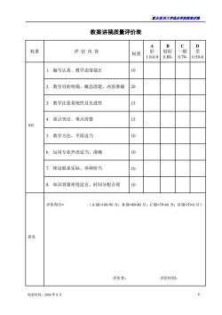
重庆医科大学临床学院教案讲稿 制表时间:2004 年 8 月 8 教案讲稿质量评价表 权重 评 估 内 容 权重 A 好 1.0-0.9 B 较好 0.89- C 一般 0.79- D 差 0.59-0 100 1. 编写认真、教学态度端正 10 2. 教学目的明确、概念清楚、内容准确 20 3. 教学注意系统性及先进性 15 4. 重点突出、难点清楚 15 5. 教学方法、手段适当 10 6. 运用专业外语适当、准确 10 7. 理论联系实际、举例恰当 10 8. 知识容量密度适宜、时间分配合理 10 意见 评价得分= (A 级=100-90 分;B 级=89-80 分;C 级=79-60 分;D 级=59-0 分) 评价者: 评价时间:
按次数下载不扣除下载券;
注册用户24小时内重复下载只扣除一次;
顺序:VIP每日次数-->可用次数-->下载券;
- 重庆医科大学:《诊断学》课程教学资源(授课教案)09 病历与诊断.doc
- 重庆医科大学:《诊断学》课程教学资源(授课教案)03 呕血.doc
- 重庆医科大学:《诊断学》课程教学资源(授课教案)04 便血.doc
- 重庆医科大学:《实验诊断学》课程教学资源(PPT课件)第八讲 脑脊液常规及生殖系统检查.ppt
- 重庆医科大学:《实验诊断学》课程教学资源(PPT课件)第七讲 大便常规、免疫学检查及心肌标志物检查.ppt
- 重庆医科大学:《实验诊断学》课程教学资源(PPT课件)第六讲 肾功能检查(主讲:唐敏).ppt
- 重庆医科大学:《实验诊断学》课程教学资源(PPT课件)第四讲 血栓与出血检查(主讲:胥文春).ppt
- 重庆医科大学:《实验诊断学》课程教学资源(PPT课件)第三讲 骨髓细胞学检查、血型与输血.ppt
- 重庆医科大学:《实验诊断学》课程教学资源(PPT课件)第一讲 总论及血液一般检查(上).ppt
- 上海科学技术出版社:《实验诊断学——彩色图谱》书籍PDF电子版(主编:张丽霞、陈金宝).pdf
- 重庆医科大学:《实验诊断学》课程复习题集(无答案).doc
- 《实验诊断学》课程实验指导书 laboatoty diagnosis(共九章,含练习及答案).doc
- 重庆医科大学:《实验诊断学》课程教学资源(教案)第8讲 CSF生殖系统.doc
- 重庆医科大学:《实验诊断学》课程教学资源(教案)第7讲 大便 免疫 心肌.doc
- 重庆医科大学:《实验诊断学》课程教学资源(教案)第6讲 肾功能.doc
- 重庆医科大学:《实验诊断学》课程教学资源(教案)第2讲 血液(下).doc
- 重庆医科大学:《实验诊断学》课程实验教学大纲(供临床医学专业本科用).doc
- 重庆医科大学:《实验诊断学》课程理论教学大纲(供五年制临床医学、儿科医学、预防医学、麻醉医学、影像医学专业本科使用).doc
- 石河子大学:《妇产科护理学》课程教学课件(PPT讲稿)第四章 妊娠期妇女的护理.ppt
- 石河子大学:《妇产科护理学》课程教学课件(PPT讲稿)第二章 女性生殖系统解剖与生理.ppt
- 重庆医科大学:《诊断学》课程教学资源(授课教案)01 绪论与问诊.doc
- 重庆医科大学:《诊断学》课程教学资源(授课教案)02 水肿.doc
- 重庆医科大学:《诊断学》课程教学资源(授课教案)07 黄疸.doc
- 重庆医科大学:《诊断学》课程教学资源(授课教案)05 腹痛.doc
- 重庆医科大学:《诊断学》课程教学资源(授课教案)06 腹泻.doc
- 重庆医科大学:《诊断学》课程教学资源(授课教案)14 心电图.doc
- 重庆医科大学:《诊断学》课程教学资源(授课教案)11 心脏检查.doc
- 重庆医科大学:《诊断学》课程教学资源(授课教案)20 呼吸系统症状和体征.doc
- 重庆医科大学:《诊断学》课程教学资源(授课教案)13 循环系统主要症状与体征.doc
- 重庆医科大学:《诊断学》课程教学资源(授课教案)15 超声心动图检查.doc
- 重庆医科大学:《诊断学》课程教学资源(授课教案)12 血管检查.doc
- 重庆医科大学:《诊断学》课程教学资源(授课教案)10 发热.doc
- 重庆医科大学:《诊断学》课程教学资源(授课教案)19 胸部检查.doc
- 重庆医科大学:《诊断学》课程教学资源(授课教案)18 呼吸困难.doc
- 重庆医科大学:《诊断学》课程教学资源(授课教案)16 咳嗽、咳痰.doc
- 重庆医科大学:《诊断学》课程教学资源(授课教案)17 咯血.doc
- 医学临床本科《MRI诊断学》课程教学大纲 Magnetic Resonance Imaging Diagnosis.doc
- 医学临床本科《影像诊断学》课程教学大纲 medical imaging(包含MRI部分).doc
- 医学影像本科《MRI诊断学》课程教学大纲(Magnetic Resonance Imaging Diagnosis).doc
- 石河子大学:《MRI诊断学》课程教学资源(讲稿,共五章).doc
