同济大学:《人体解剖学 Human anatomy》课程电子教案(PPT课件)ABDOMEN Ⅳ

ABDOMEN IV liyingchd@163.com
ABDOMEN Ⅳ liyingchd@163.com
Objects Go over the stomach and the liver The arteries and ligaments of stomach and liver ·Bile circulation ·Pancreas ·Small intestine ·Large intestine Dissection 3
Objects 1 • Go over the stomach and the liver • The arteries and ligaments of stomach and liver 2 • Bile circulation • Pancreas • Small intestine • Large intestine • 3 •Dissection
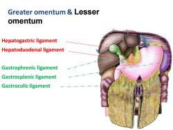
Greater omentum Lesser omentum Hepatogastric ligament Hepatoduodenal ligament Gastrophrenic ligament Gastrosplenic ligament Gastrocolic ligament
Hepatogastric ligament Hepatoduodenal ligament Gastrophrenic ligament Gastrosplenic ligament Gastrocolic ligament Greater omentum & Lesser omentum
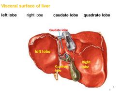
Visceral surface of liver left lobe right lobe caudate lobe quadrate lobe Caudate lobe left lobe Right lobe
Visceral surface of liver left lobe right lobe caudate lobe quadrate lobe 4 Right Quadrate lobe lobe Caudate lobe left lobe

Liver *transverse fissure--Porta hepatis 5 cm long,transversely across the "H" -Contents Right and left hepatic ducts Left and right branches of proper hepatic artery Left and right branches of hepatic portal vein Nerves and lymphatic vessels These structures are surrounded by connective tissue and is called “hepatic pedicle
Liver ★ transverse fissure -- Porta hepatis 5 cm long, transversely across the “H”, – Contents • Right and left hepatic ducts • Left and right branches of proper hepatic artery • Left and right branches of hepatic portal vein • Nerves and lymphatic vessels – These structures are surrounded by connective tissue and is called “hepatic pedicle
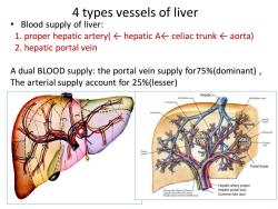
4 types vessels of liver Blood supply of liver: 1.proper hepatic artery(←hepatic A←celiac trunk←aorta) 2.hepatic portal vein A dual BLOOD supply:the portal vein supply for75%(dominant), The arterial supply account for 25%(lesser) Hepatic v. Sublobular vei Sublobular veins Portal triads Hepatic artery proper Hepatic portal vein ptisnand bie duct Common bile duct
• Blood supply of liver: 1. proper hepatic artery( ← hepatic A← celiac trunk ← aorta) 2. hepatic portal vein A dual BLOOD supply: the portal vein supply for75%(dominant) , The arterial supply account for 25%(lesser) 4 types vessels of liver

1.Proper hepatic artery Celiac trunk→common hepatic A→proper hepatic A>right left branches
1. Proper hepatic artery Celiac trunk → common hepatic A →proper hepatic A → right & left branches →

2.Hepatic portal vein General features the union of superior mesenteric vein SMV and splenic vein passing through the lesser omentum to the porta hepatis,divides into right and left branches Has no valves in hepatic portal system Drains blood from GI tract(capillaries) to hepatic sinusoid(capillaries):the lower end of esophagus to the upper end of anal canal,pancreas,gall bladder, bile ducts and spleen
2. Hepatic portal vein General features • the union of superior mesenteric vein SMV and splenic vein • passing through the lesser omentum to the porta hepatis, divides into right and left branches • Has no valves in hepatic portal system • Drains blood from GI tract(capillaries) to hepatic sinusoid(capillaries) : the lower end of esophagus to the upper end of anal canal, pancreas, gall bladder, bile ducts and spleen
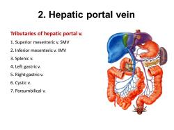
2.Hepatic portal vein Tributaries of hepatic portal v. 1.Superior mesenteric v.SMV 2.Inferior mesenteric v.IMV 3.Splenic v. 4.Left gastricv. 5.Right gastric v. 6.Cystic v. 7.Paraumbilical v
2. Hepatic portal vein Tributaries of hepatic portal v. 1. Superior mesenteric v. SMV 2. Inferior mesenteric v. IMV 3. Splenic v. 4. Left gastric v. 5. Right gastric v. 6. Cystic v. 7. Paraumbilical v

Portal-Caval venous anastomoses 1.Site at the esophagus left gastric vein→esophageal venous plexus→ esophageal vein>hemiazygos vein superior vena cava Esophageal varices,---bleeding 2.At rectum splenic vein>inferior mesenteric vein>superior rectal vein>rectal venous plexus>inferior rectal and anal veins>internal iliac vein>inferior vena cava Hemorroid ---Hemorrage Portal hypertension
Portal- Caval venous anastomoses 1. Site at the esophagus left gastric vein → esophageal venous plexus → esophageal vein → hemiazygos vein → superior vena cava Esophageal varices,---bleeding 2. At rectum splenic vein → inferior mesenteric vein → superior rectal vein → rectal venous plexus→ inferior rectal and anal veins → internal iliac vein → inferior vena cava • Hemorroid --- Hemorrage Portal hypertension
按次数下载不扣除下载券;
注册用户24小时内重复下载只扣除一次;
顺序:VIP每日次数-->可用次数-->下载券;
- 同济大学:《人体解剖学 Human anatomy》课程电子教案(PPT课件)The abdomen(三).pptx
- 同济大学:《人体解剖学 Human anatomy》课程电子教案(PPT课件)The abdomen(二).ppt
- 同济大学:《人体解剖学 Human anatomy》课程电子教案(PPT课件)HUMAN ANATOMY.ppt
- 同济大学:《人体解剖学 Human anatomy》课程电子教案(PPT课件)Skull.pdf
- 同济大学:《人体解剖学 Human anatomy》课程电子教案(PPT课件)Brain’s blood supply.pdf
- 同济大学:《人体解剖学 Human anatomy》课程电子教案(PPT课件)The Spinal Cord.pdf
- 同济大学:《人体解剖学 Human anatomy》课程电子教案(PPT课件)Cranial Nerves.pdf
- 同济大学:《人体解剖学 Human anatomy》课程电子教案(PPT课件)Brainstem.pdf
- 同济大学:《人体解剖学 Human anatomy》课程电子教案(PPT课件)Brainstem.pdf
- 同济大学:《人体解剖学 Human anatomy》课程电子教案(PPT课件)ELBOW FOREARM WRIST & HAND.ppt
- 同济大学:《人体解剖学 Human anatomy》课程电子教案(PPT课件)Shoulder & Arm.ppt
- 同济大学:《人体解剖学 Human anatomy》课程电子教案(PPT课件)The Female Reproductive System.ppt
- 同济大学:《人体解剖学 Human anatomy》课程电子教案(PPT课件)The Cardiovascular System.ppt
- 同济大学:《人体解剖学 Human anatomy》课程电子教案(PPT课件)The Heart.ppt
- 同济大学:《人体解剖学 Human anatomy》课程电子教案(PPT课件)Deep Structures in the Cervical Region.ppt
- 同济大学:《人体解剖学 Human anatomy》课程电子教案(PPT课件)Bones of Upper Limb.pdf
- 同济大学:《人体解剖学 Human anatomy》课程电子教案(PPT课件)Telencephalon.pdf
- 同济大学:《人体解剖学 Human anatomy》课程电子教案(PPT课件)Cerebellum.pdf
- 同济大学:《人体解剖学 Human anatomy》课程电子教案(PPT课件)Anterior and Medial Thigh.pdf
- 同济大学:《人体解剖学 Human anatomy》课程电子教案(PPT课件)The Cervical Region.pdf
- 同济大学:《人体解剖学 Human anatomy》课程电子教案(PPT课件)Abdomen Ⅴ.ppt
- 同济大学:《人体解剖学 Human anatomy》课程电子教案(PPT课件)Regions and Triangles of the Neck.ppt
- 同济大学:《人体解剖学 Human anatomy》课程电子教案(PPT课件)Mediastinum.ppt
- 同济大学:《人体解剖学 Human anatomy》课程电子教案(PPT课件)SPLANCHNOLOGY.ppt
- 同济大学:《人体解剖学 Human anatomy》课程电子教案(PPT课件)The Cervical Region.ppt
- 同济大学:《人体解剖学 Human anatomy》课程电子教案(PPT课件)Deep Structures in the Cervical Region.ppt
- 同济大学:《人体解剖学 Human anatomy》课程电子教案(PPT课件)Visual Organ.ppt
- 同济大学:《人体解剖学 Human anatomy》课程电子教案(PPT课件)Pelvis.ppt
- 郑州大学:儿科医学知识图谱命名实体和关系标注规范(讲义).pdf
- 郑州大学:《心电图学》课程教学资源(课件讲稿)第三讲 心电图一般知识(李中健、李世锋、申继红、刘儒).pdf
- 郑州大学:《心电图学》课程教学资源(课件讲稿)心电现象之coumel定律(主讲:成媛).pdf
- 郑州大学:《心电图学》课程教学资源(课件讲稿)第十三讲 宽QRS波心动过速.pdf
- 郑州大学:《心电图学》课程教学资源(课件讲稿)第五讲 心电图各波段正常值.pdf
- 基础医学(学习资料)易制毒化学品分类和品种目录.doc
- 基础医学(学习资料)易制爆危险化学品名录(2017年版).doc
- 基础医学(学习资料)剧毒化学品目录(2015版).docx
- 郑州大学:《微生物学与免疫学》课程教学资源(课件讲义)免疫学新进展(New advance in Immunology)教学大纲.docx
- 郑州大学:《微生物学与免疫学》课程教学资源(课件讲义)自身免疫性疾病 Autoimmune Disease.docx
- 郑州大学:《微生物学与免疫学》课程教学资源(课件讲义)免疫学教案(任课教师:杜英).docx
- 郑州大学:《微生物学与免疫学》课程教学资源(课件讲义)免疫学基础(打印版)Medical Immunology.docx
