重庆医科大学:《内科学》课程教学课件(PPT讲稿)Pericardial diease
Pericardial diease Pericarditis
Pericardial diease Pericarditis
Introduction.The patterns of pericardial disease have changed over the last 20-30 years,with the once common tuberculous pericarditis becoming a rare entity.Uremic pericarditis formerly one of the most dreaded complications of renal failure,is now managed with relative ease. Pericarditis can be acute and self-limited,subacute, or chronic.Chronic forms of pericarditis often result in cardiac constriction.A variety of agents cause pericarditis.Pericarditis is likely to lead to effusion, which may be minor or large enough to interfere with ventricular filling during diastole (tamponade). Chronic forms of pericarditis often are associated with fibrosis,a condition that can also impede diastolic,ventricular filling
• Introduction. The patterns of pericardial disease have changed over the last 20-30 years, with the once common tuberculous pericarditis becoming a rare entity. Uremic pericarditis , formerly one of the most dreaded complications of renal failure, is now managed with relative ease. Pericarditis can be acute and self-limited, subacute, or chronic. Chronic forms of pericarditis often result in cardiac constriction. A variety of agents cause pericarditis. Pericarditis is likely to lead to effusion, which may be minor or large enough to interfere with ventricular filling during diastole (tamponade). Chronic forms of pericarditis often are associated with fibrosis, a condition that can also impede diastolic, ventricular filling
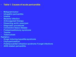
Table 1.Causes of acute pericarditis Malignant tumor Idiopathic pericarditis Uremia Bacteria infection Anticoagulant therapy Dissecting aortic aneurysm Diagnostic procedures Connective tissue disease Postpericardiotomy syndrome Trauma Tuberculosis Radiation Drugs inducing lupuslike syndrome Chylopericardium Postmyocardial infraction syndrome Fungal infections AlDS-related pericarditis
• Table 1. Causes of acute pericarditis • Malignant tumor • Idiopathic pericarditis • Uremia • Bacteria infection • Anticoagulant therapy • Dissecting aortic aneurysm • Diagnostic procedures • Connective tissue disease • Postpericardiotomy syndrome • Trauma • Tuberculosis Radiation • Drugs inducing lupuslike syndrome • Chylopericardium • Postmyocardial infraction syndrome Fungal infections • AIDS-related pericarditis
Pathology 。The histopathology of the acute pericarditis is dependent on the underlying etiology.The majority of cases are manifest by hyperemia and increased microvascularity, polymorpholeukocyte accumulation and fibrin deposition. Adhesions can form between the layers of the pericardium,and between pericardium and adjacent structure such as pleura and mediastinum
Pathology • The histopathology of the acute pericarditis is dependent on the underlying etiology. The majority of cases are manifest by hyperemia and increased microvascularity, polymorpholeukocyte accumulation and fibrin deposition. Adhesions can form between the layers of the pericardium, and between pericardium and adjacent structure such as pleura and mediastinum
● History.Much of the historical information obtain from patients with pericarditis varies according to the underlying medical condition.Thus,rheumatoid pericarditis usually accompanies severe rheumatoid arthritis,while radiation pericarditis follows large doses of mediastinal radiation, ordinarily administerd for control of neoplasm.Systemic symptoms may occur,including fever,sweats,chills. weakness,malaise,weight loss,anxiety,and depression.The pain of pericarditis is characteristic: It is almost always precordial in position,sharp and severe,and increasing in severity with deep inspiration or recumbency.Occasionally,pain is felt only when patient lie on the left side and make a maximum inspiratory effort.The pain not infrequently radiates to the neck or left arm and shoulder. Dyspnea usually is seen only in individuals with cardiac tamponade or constriction.Palpitations are noted commonly.These are caused by atrial arrhythmias,which often accompany pericarditis
• History. Much of the historical information obtain from patients with pericarditis varies according to the underlying medical condition. Thus, rheumatoid pericarditis usually accompanies severe rheumatoid arthritis, while radiation pericarditis follows large doses of mediastinal radiation, ordinarily administerd for control of neoplasm. Systemic symptoms may occur, including fever, sweats, chills, weakness, malaise, weight loss, anxiety, and depression. The pain of pericarditis is characteristic: It is almost always precordial in position, sharp and severe, and increasing in severity with deep inspiration or recumbency. Occasionally, pain is felt only when patient lie on the left side and make a maximum inspiratory effort. The pain not infrequently radiates to the neck or left arm and shoulder. Dyspnea usually is seen only in individuals with cardiac tamponade or constriction. Palpitations are noted commonly. These are caused by atrial arrhythmias, which often accompany pericarditis
Physical examination.Patients with pericarditis may have an irregular or rapid pulse secondary to atrial arrhythmias or tamponade.A pericardial friction rub is the hallmark of the disease.This superficial scraping, scratchy,or grating sound is frequently evanescent or intermittent.Persistent rubs are most often heard with Pericarditis secondary to neoplasm,uremia,viral infection,and tuberculosis.Evanescent rubs are common in pericarditis associated with Ml.Rubs may have one,two,or three components occurring with the following stages of cardiac cycle:(1)rapid filling in early diastole,(2)atrial contraction in late diastole,and (3) systole.On occasion,one-or two-component rubs are confused with systolic or to-and-fro murmurs.Sometimes a patient with pericarditis has an early diastolic sound or pericardial knock,which can be confused with a third heart sound (S3).Other systemic findings in patients with pericarditis depend on the underlying condition
• Physical examination. Patients with pericarditis may have an irregular or rapid pulse secondary to atrial arrhythmias or tamponade. A pericardial friction rub is the hallmark of the disease. This superficial scraping, scratchy, or grating sound is frequently evanescent or intermittent. Persistent rubs are most often heard with Pericarditis secondary to neoplasm, uremia, viral infection, and tuberculosis. Evanescent rubs are common in pericarditis associated with MI. Rubs may have one, two, or three components occurring with the following stages of cardiac cycle: (1) rapid filling in early diastole, (2) atrial contraction in late diastole, and (3) systole. On occasion, one- or two-component rubs are confused with systolic or to-and-fro murmurs. Sometimes a patient with pericarditis has an early diastolic sound or pericardial knock, which can be confused with a third heart sound (S3). Other systemic findings in patients with pericarditis depend on the underlying condition
ECG.The ECG is often abnormal in patients with pericarditis.ECG findings include diffusely elevated S-T segments,widespread T wave changes, and P-R segment depression.Elevated S-T segments can mimic the ECG findings of acute Ml.ECG findings in pericarditis are as follows: Elevation of S-T segment Flattening and inversion of T waves Depression of P-R segment
• ECG. The ECG is often abnormal in patients with pericarditis. ECG findings include diffusely elevated S-T segments, widespread T wave changes, and P-R segment depression. Elevated S-T segments can mimic the ECG findings of acute MI. ECG findings in pericarditis are as follows: Elevation of S-T segment Flattening and inversion of T waves Depression of P-R segment
Their differentiation from ECG changes of acute MI are listed in Table 2.The S-T segment elevations of early repolarization occasionally may be confused with those of pericarditis; however,S-T segment elevation of early repolarization usually is limited to the precordial leads,while that of pericarditis occurs throughout limb and precordial leads.Supraventricular arrhythmias are frequently noted in patients with pericarditis
• Their differentiation from ECG changes of acute MI are listed in Table 2. The S-T segment elevations of early repolarization occasionally may be confused with those of pericarditis; however, S-T segment elevation of early repolarization usually is limited to the precordial leads, while that of pericarditis occurs throughout limb and precordial leads. Supraventricular arrhythmias are frequently noted in patients with pericarditis

Figure 1 Electrocardiographic recording of a 50-year-old man in sinus tachycardia with T wave abnormality and findings of stage 1 pericarditis
• Figure 1 Electrocardiographic recording of a 50-year-old man in sinus tachycardia with T wave abnormality and findings of stage 1 pericarditis
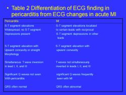
Table 2 Differentiation of ECG finding in pericarditis from ECG changes in acute MI Pericarditis MI S-T segment elevations S-T segment elevations localized Widespread;no S-T segment to certain leads with reciprocal Depressions present S-T segment depressions in other leads S-T segment elevation with S-T segment elevation with Upward concavity or straight upward convexity Morphology Simultaneous T wave inversion T waves not simultaneously In lead I,Il,and Ill inverted in leads 1,Il,and Ill Significant Q waves not seen significant Q waves frequently With pericarditis seen with MI QRS often normal QRS often abnormal
• Table 2 Differentiation of ECG finding in pericarditis from ECG changes in acute MI Pericarditis MI S-T segment elevations S-T segment elevations localized Widespread; no S-T segment to certain leads with reciprocal Depressions present S-T segment depressions in other leads S-T segment elevation with S-T segment elevation with Upward concavity or straight upward convexity Morphology Simultaneous T wave inversion T waves not simultaneously In lead I, II, and III inverted in leads I, II, and III Significant Q waves not seen significant Q waves frequently With pericarditis seen with MI QRS often normal QRS often abnormal
按次数下载不扣除下载券;
注册用户24小时内重复下载只扣除一次;
顺序:VIP每日次数-->可用次数-->下载券;
- 重庆医科大学:《内科学》课程教学课件(PPT讲稿)Leukemia(一).ppt
- 重庆医科大学:《内科学》课程教学课件(PPT讲稿)hypertension.ppt
- 重庆医科大学:《内科学》课程教学课件(PPT讲稿)HEART FAILURE.ppt
- 重庆医科大学:《内科学》课程教学课件(PPT讲稿)Glomerulardisease.ppt
- 重庆医科大学:《内科学》课程教学课件(PPT讲稿)Generalintroductionofrenaldisease.ppt
- 重庆医科大学:《内科学》课程教学课件(PPT讲稿)Chronic Renal Failure.ppt
- 重庆医科大学:《内科学》课程教学课件(PPT讲稿)Cardiomyopathies.ppt
- 重庆医科大学:《内科学》课程教学课件(PPT讲稿)高血压病 PRIMARY HYPERTENTION.ppt
- 重庆医科大学:《内科学》课程教学课件(PPT讲稿)心肌病(附属第二医院心内科:高大中).ppt
- 重庆医科大学:《内科学》课程教学课件(PPT讲稿)心脏瓣膜病(valvular heart disease).ppt
- 重庆医科大学:《内科学》课程教学课件(PPT讲稿)心肌炎(myocarditis).ppt
- 重庆医科大学:《内科学》课程授课教案(七年制)贫血总论 - 陈姝.doc
- 重庆医科大学:《内科学》课程授课教案(七年制)血小板减少性紫癜 - 邓建川.doc
- 重庆医科大学:《内科学》课程授课教案(七年制)胃食管反流病 - 冯晓霞.doc
- 重庆医科大学:《内科学》课程授课教案(七年制)胃炎 - 宁波.doc
- 重庆医科大学:《内科学》课程授课教案(七年制)肾上腺皮质功能亢进 - 程伟.doc
- 重庆医科大学:《内科学》课程授课教案(七年制)肝硬化 - 沈薇.doc
- 重庆医科大学:《内科学》课程授课教案(七年制)肝性脑病 - 沈薇.doc
- 重庆医科大学:《内科学》课程授课教案(七年制)缺铁性贫血 - 陈姝.doc
- 重庆医科大学:《内科学》课程授课教案(七年制)糖尿病 - 龙敏.doc
- 重庆医科大学:《内科学》课程教学课件(PPT讲稿)系统性红斑狼疮狼疮性肾炎(Systemic Lupus Erythematosus,SLE).ppt
- 重庆医科大学:《内科学》课程教学课件(PPT讲稿)冠状动脉粥样硬化型心脏病 Coronary Atherosclerotic Heart Disease.ppt
- 重庆医科大学:《内科学》课程教学课件(PPT讲稿)心律失常 Cardiac Arrhythmia.ppt
- 重庆医科大学:《内科学》课程教学课件(PPT讲稿)Leukemia(二).ppt
- 重庆医科大学:《内科学》课程教学课件(PPT讲稿)再生障碍性贫血 Aplastic Anemia.ppt
- 重庆医科大学:《内科学》课程教学课件(PPT讲稿)功能性胃肠病 Functional Gastrointestinal Disorders.ppt
- 重庆医科大学:《内科学》课程教学课件(PPT讲稿)第七篇 内分泌和代谢疾病 第一章 总论.ppt
- 重庆医科大学:《内科学》课程教学课件(PPT讲稿)第七篇 内分泌和代谢疾病 第九章 甲状腺功能亢进症 Hyperthyroidism.ppt
- 重庆医科大学:《内科学》课程教学课件(PPT讲稿)第七篇 内分泌和代谢疾病 第六章 尿崩症.ppt
- 重庆医科大学:《内科学》课程教学课件(PPT讲稿)第七篇 内分泌和代谢疾病 第十三章 库欣综合征(Cushing’s syndrome).ppt
- 重庆医科大学:《内科学》课程教学课件(PPT讲稿)第七篇 内分泌和代谢疾病 第十六章 嗜铬细胞瘤.ppt
- 重庆医科大学:《内科学》课程教学课件(PPT讲稿)第七篇 内分泌和代谢疾病 第十四章 原发性醛固酮增多症.ppt
- 重庆医科大学:《内科学》课程教学课件(PPT讲稿)糖尿病.ppt
- 重庆医科大学:《内科学》课程教学课件(PPT讲稿)缺铁性贫血 Iron deficiency anemia.ppt
- 《运动解剖学》课程文献资料(脑科学)中国科学院脑科学与智能技术卓越创新中心非人灵长类研究平台.pdf
- 《运动解剖学》课程文献资料(脑科学)中国脑科学计划进展.pdf
- 《运动解剖学》课程文献资料(脑科学)农村小学科学课程资源对教学效果的影响研究.pdf
- 《运动解剖学》课程文献资料(脑科学)双减背景下六步项目化”学习在初中科学教学中的研究.pdf
- 《运动解剖学》课程文献资料(脑科学)学习科学与科学教育的共同演——与国际学习科学学会前主席马西娅·林教授对话.pdf
- 《运动解剖学》课程文献资料(脑科学)学习科学的内涵、价值特征及其对课堂教学的指引_陈娜.pdf
