同济大学:《眼科学》课程电子教案(PPT课件讲稿)视网膜疾病 Retinal Disease

Retinal Disease Jingfa Zhang,Ph.D. Tongji Eye Institute,Tongji University School of Medicine
Retinal Disease Jingfa Zhang, Ph.D. Tongji Eye Institute, Tongji University School of Medicine
Outline Anatomy of the eye and the retina ● Retinal diagnostic test Diabetic Retinopathy (DR) Age-related Macular Degeneration (AMD)
• Anatomy of the eye and the retina • Retinal diagnostic test • Diabetic Retinopathy (DR) • Age-related Macular Degeneration (AMD) Outline

Outline Anatomy of the eye and the retina ● Retinal diagnostic test Diabetic Retinopathy (DR) Age-related Macular Degeneration (AMD)
• Anatomy of the eye and the retina • Retinal diagnostic test • Diabetic Retinopathy (DR) • Age-related Macular Degeneration (AMD) Outline
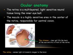
Ocular anatomy The retina is a multilayered,light sensitive neural tissue lining the inner eye ball. The macula is a highly sensitive area in the center of the retina,responsible for central vision. RETINA SCLERA NS -OVEA IRIS MACULA CHOROID The vitreous -clear gel fills the back of the eye and sits in front of the retina OPTIC NERVE CORNEA The retina -senses light transmits images to the brain
• The retina is a multilayered, light sensitive neural tissue lining the inner eye ball. • The macula is a highly sensitive area in the center of the retina, responsible for central vision. Ocular anatomy The vitreous – clear gel fills the back of the eye and sits in front of the retina The retina – senses light & transmits images to the brain
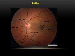
Retina Optic Cup Fovea Optic Disc Macula Artery Vein Stanford Medidine 5
Retina
Retinal histology ·Ten layers ·Four cell types: ·Wacula/.Fovea ·Optic nerve head
Retinal histology • Ten layers • Four cell types: • Macula/Fovea • Optic nerve head

Retinal Layers Cells NFL Nerve fiber layer Inner limiting membrane GCL Ganglion cell Axons at surface of layer retina passing via optic nerve,chiasm and tract to lateral IPL Inner plexiform geniculate body layer Ganglion cell INL Inner nuclear Muller cell layer (supporting glial cell) Bipolar cell OPL Outer plexiform Amarcine cell layer Horizontal cell Rod ONL Outer nuclear Cone layer Pigment cells OLM of choroid RCL Photoreceptor layer Pigment B.Section RPE epithelium through retina
ILM NFL GCL IPL INL OPL ONL OLM RCL RPE
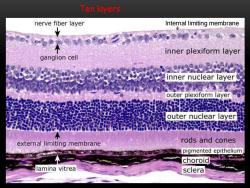
Ten layers nerve fiber layer Internal limiting membrane inner plexiform layer ganglion cell inner nuclear layer outer plexiform layer 8吧 outer nuclear layer external limiting membrane rods and cones pigmented epithelium choroid lamina vitrea sclera
Ten layers Internal limiting membrane
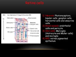
Retina cells S254 Surv Ophthalmol 47(Suppl 2)December 2002 1)Neurons:Photoreceptors, Glial cells Blood vessels hv bipolar cells,ganglion cells, horizontal cells and amacrine Astrocytes Neurons cells Ganglion cells 2)Blood vessels:endothelial Muller cells Bipolar cells cells and pericyte Horizontal cells Microglia 3)Glial cells:Marcoglia Amacrine cells (Astrocyte and Muller cells) Photoreceptors and microglia 4)RPE:retinal pigmented epithelium
Retina cells 1) Neurons: Photoreceptors, bipolar cells, ganglion cells, horizontal cells and amacrine cells 2) Blood vessels: endothelial cells and pericyte 3) Glial cells: Marcoglia (Astrocyte and Muller cells) and microglia 4) RPE: retinal pigmented epithelium
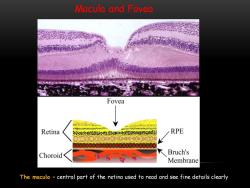
Macula and Fovea Fovea 4- Retina RPE Choroid Bruch's Membrane The macula central part of the retina used to read and see fine details clearly
Macula and Fovea The macula – central part of the retina used to read and see fine details clearly
按次数下载不扣除下载券;
注册用户24小时内重复下载只扣除一次;
顺序:VIP每日次数-->可用次数-->下载券;
- 同济大学:《眼科学》课程电子教案(PPT课件讲稿)视光学基础 Basic Optics.ppt
- 同济大学:《眼科学》课程电子教案(PPT课件讲稿)葡萄膜炎 Disease of the Uvea.ppt
- 同济大学:《眼科学》课程电子教案(PPT课件讲稿)眼组织学 Histology of the Eye.ppt
- 同济大学:《眼科学》课程电子教案(PPT课件讲稿)眼科导论 Introduction(负责人:徐国彤).ppt
- 同济大学:《眼科学》课程教学资源(大纲教案)眼科学教学大纲(英文,Ophthalmology).doc
- 同济大学:《眼科学》课程教学资源(大纲教案)眼科学教学大纲(中文,负责人:徐国彤).doc
- 同济大学:《眼科学》课程电子教案(PPT课件讲稿)白内障 Ophthalmology.ppt
- 同济大学:《眼科学》课程电子教案(PPT课件讲稿)眼睑和眼表疾病 The eyelids & ocular surface diseases.ppt
- 同济大学:《眼科学》课程电子教案(PPT课件讲稿)眼眶异常 Disorders of the orbit.ppt
- 同济大学:《眼科学》课程电子教案(PPT课件讲稿)斜视 STRABISMUS.ppt
- 同济大学:《眼科学》课程电子教案(PPT课件讲稿)巩膜病 Scleral Disease.ppt
- 同济大学:《眼科学》课程电子教案(PPT课件讲稿)屈光学 Optics and Refractive errors correction.ppt
- 同济大学:《眼科学》课程电子教案(PPT课件讲稿)屈光不正 Our eye as a camera Refraction, errors and solutions..ppt
- 同济大学:《眼科学》课程电子教案(PPT课件讲稿)泪道疾病 THE WATERING EYE.ppt
- 同济大学:《眼科学》课程教学资源(试卷习题)MBBS试题.doc
- 同济大学:《眼科学》课程教学资源(试卷习题)MBBS眼科试卷-B.doc
- 同济大学:《眼科学》课程教学资源(试卷习题)MBBS眼科试卷-A.doc
- 《医学免疫学》课程教学资源(参考资料)医学免疫学常见问题解答.docx
- 广东医科大学:《医学免疫学》课程教学资源(打印版)教学大纲 Medical Immunology(负责人:米娜).pdf
- 《医学免疫学》课程教学资源(参考资料)医学免疫学相关词汇合集.docx
- 同济大学:《眼科学》课程电子教案(PPT课件讲稿)视觉系统 Visual Organ.ppt
- 同济大学:《眼科学》课程电子教案(PPT课件讲稿)角膜病总论 Corneal disease.ppt
- 同济大学:《眼科学》课程电子教案(PPT课件讲稿)青光眼 Glaucoma.pptx
- 同济大学:《眼科学》课程电子教案(PPT课件讲稿)眼肿瘤 Ocular trauma.ppt
- 同济大学:《病理学》课程教学资源(试卷习题,含答案)Cell Injury.pdf
- 同济大学:《病理学》课程教学资源(试卷习题,含答案)Inflammation.pdf
- 同济大学:《病理学》课程教学资源(试卷习题,含答案)Neoplasia.pdf
- 同济大学:《病理学》课程教学资源(试卷习题,含答案)Thrombosis I-II, Hemodynamics, Atherosclerosis.pdf
- 同济大学:《病理学》课程教学资源(试卷习题,含答案)Cardiac pathology.pdf
- 同济大学:《病理学》课程教学资源(试卷习题,含答案)Female reproductivepathology.pdf
- 同济大学:《病理学》课程教学资源(试卷习题,含答案)GIpathology.pdf
- 同济大学:《病理学》课程教学资源(试卷习题,含答案)试题A卷.pdf
- 同济大学:《病理学》课程教学资源(试卷习题,含答案)试题B卷.pdf
- 同济大学:《病理学》课程教学资源(教案讲义)Introduction to Pathology.pdf
- 同济大学:《病理学》课程教学资源(教案讲义)Chapter 01 Adaptation and Injury of Cell and Tissue(Adaptation of Cell and Tissue).pdf
- 同济大学:《病理学》课程教学资源(教案讲义)Chapter 01 Adaptation and Injury of Cell and Tissue(Reversible Injury of Cell and Tissue).pdf
- 同济大学:《病理学》课程教学资源(教案讲义)Chapter 01 Adaptation and Injury of Cell and Tissue(Irreversible Injury of Cell and Tissue).pdf
- 同济大学:《病理学》课程教学资源(教案讲义)Chapter 02 组织再生与修复 Tissue regeneration and repair.pdf
- 同济大学:《病理学》课程教学资源(教案讲义)Chapter 14 女性生殖系统和乳房疾病 THE DISEASE OF FEMALE GENITAL SYSTEM AND BREAST.pdf
- 同济大学:《病理学》课程教学资源(教案讲义)Chapter 16 神经系统疾病 Diseases of the Nervous System.pdf
