同济大学:《眼科学》课程电子教案(PPT课件讲稿)泪道疾病 THE WATERING EYE

THE WATERING EYE Dr.JUN ZOU Shanghai Tenth People's Hospital 2016-3-9 .Ljubljana,Slovenia Eye Hospital,University Medical Centre
THE WATERING EYE Dr. JUN ZOU Shanghai Tenth People's Hospital 2016-3-9 •Ljubljana, Slovenia Eye Hospital, University Medical Centre
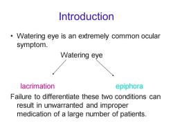
Introduction Watering eye is an extremely common ocular symptom. Watering eye lacrimation epiphora Failure to differentiate these two conditions can result in unwarranted and improper medication of a large number of patients
Introduction • Watering eye is an extremely common ocular symptom. Watering eye lacrimation epiphora Failure to differentiate these two conditions can result in unwarranted and improper medication of a large number of patients
THE WATERING EYE Lacrimation is watering that occurs secondary to excessive tear production in the presence of a normal excretory system. Epiphora is watering that occurs secondary to abnormal excretory system in the presence of normal tear secretion
THE WATERING EYE • Lacrimation is watering that occurs secondary to excessive tear production in the presence of a normal excretory system. • Epiphora is watering that occurs secondary to abnormal excretory system in the presence of normal tear secretion
Secretory system Lacrimal gland:orbital part(2/3) palpebral part(1/3) Accessory lacrimal glands:Krause Wolfring
Secretory system • Lacrimal gland: orbital part (2/3) palpebral part (1/3) • Accessory lacrimal glands: Krause Wolfring
Secretory system Normal tear secretion is necessary to keep the ocular surface moist.The tears form a thin film,which integrity is crucial for normal corneal physiology. Tear composition and secretion three layers: the mucin layer:secreted by the goblet cells;alters the surface tension of the tears and increases its adherence to the cornea. the aqueous layer:secreted by the accessory lacrimal glands (Krause and Wolfring)or in the presence of ocular irritation the main lacrimal gland. the oily layer:secreted by the Meibomian glands;prevents tear film evaporation
Secretory system • Normal tear secretion is necessary to keep the ocular surface moist. The tears form a thin film, which integrity is crucial for normal corneal physiology. • Tear composition and secretion - three layers: • the mucin layer: secreted by the goblet cells; alters the surface tension of the tears and increases its adherence to the cornea. • the aqueous layer: secreted by the accessory lacrimal glands (Krause and Wolfring) or in the presence of ocular irritation the main lacrimal gland. • the oily layer: secreted by the Meibomian glands; prevents tear film evaporation
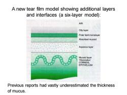
A new tear film model showing additional layers and interfaces (a six-layer model): AIR Oily layer Polar lipid monolayer Absorbed mucoid Aqueous layer Mucoid layer Glycocalyx' CORNEAL EPITHELIUM Previous reports had vastly underestimated the thickness of mucus
A new tear film model showing additional layers and interfaces (a six-layer model): Previous reports had vastly underestimated the thickness of mucus
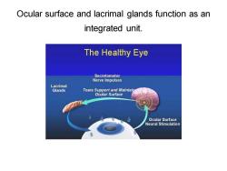
Ocular surface and lacrimal glands function as an integrated unit. The Healthy Eye Secretomotor Nerve Impulses Lacrimal Glands Tears Support and Maintaio Ocular Surface Ocular Surface Neural Stimulation
Ocular surface and lacrimal glands function as an integrated unit

Secretory system Pathology of the tear film: deficiency of the aqueous layer -keratoconjunctivitis sicca. deficiency of mucous or meibomian secretions instability of the tear film(compensatory excessive aqueous secretion -paradoxical watering)
Secretory system • Pathology of the tear film: • deficiency of the aqueous layer - keratoconjunctivitis sicca. • deficiency of mucous or meibomian secretions - instability of the tear film (compensatory excessive aqueous secretion - paradoxical watering)
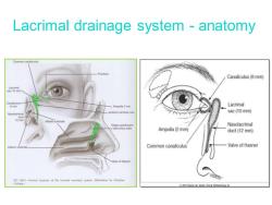
Lacrimal drainage system -anatomy Common canaliculus Punctum Canalicuus (8 mm) 8 mm -Amputls 2 mm Lacrimal sac (10 mm) 12m Middle turbinate Nasolacrimal Ampulla (2 mm duct (12 mm) Common canaloulus Valve of Hasner manmy te lcimal ecrymn byChne 2007E
Lacrimal drainage system - anatomy
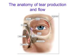
The anatomy of tear production and flow Lacrimal gland Ca Lacrimal sac Middle turbinate Nasolacrimal duct Inferior turbinate
The anatomy of tear production and flow
按次数下载不扣除下载券;
注册用户24小时内重复下载只扣除一次;
顺序:VIP每日次数-->可用次数-->下载券;
- 同济大学:《眼科学》课程教学资源(试卷习题)MBBS试题.doc
- 同济大学:《眼科学》课程教学资源(试卷习题)MBBS眼科试卷-B.doc
- 同济大学:《眼科学》课程教学资源(试卷习题)MBBS眼科试卷-A.doc
- 《医学免疫学》课程教学资源(参考资料)医学免疫学常见问题解答.docx
- 广东医科大学:《医学免疫学》课程教学资源(打印版)教学大纲 Medical Immunology(负责人:米娜).pdf
- 《医学免疫学》课程教学资源(参考资料)医学免疫学相关词汇合集.docx
- 广东医科大学:《生理学》课程教学资源(课件讲稿,打印版)第十章 内分泌 Endocrine.pdf
- 广东医科大学:《生理学》课程教学资源(课件讲稿,打印版)第十一章 生殖 Reproduction.pdf
- 广东医科大学:《生理学》课程教学资源(课件讲稿,打印版)第六章 消化和吸收 Digestion and Absorption.pdf
- 广东医科大学:《生理学》课程教学资源(课件讲稿,打印版)第八章 尿的生成和排出 Formation and excretion of the urine.pdf
- 广东医科大学:《生理学》课程教学资源(课件讲稿,打印版)第五章 呼吸 Respiration.pdf
- 广东医科大学:《生理学》课程教学资源(课件讲稿,打印版)第九章 神经系统的功能 Function of Nervous System.pdf
- 广东医科大学:《生理学》课程教学资源(课件讲稿,打印版)第四章 血液循环 Blood Circulation.pdf
- 广东医科大学:《生理学》课程教学资源(课件讲稿,打印版)第三章 血液 Blood.pdf
- 广东医科大学:《生理学》课程教学资源(课件讲稿,打印版)第二章 细胞的基本功能 Basic functions of cells.pdf
- 广东医科大学:《生理学》课程教学资源(课件讲稿,打印版)第一章 绪论 physiology(负责人:张秀娟).pdf
- 广东医科大学:《生理学》课程教学资源(讲义)教学重点与难点内容(共十一章).pdf
- 广东医科大学:《生理学》课程教学资源(资料)生理学专业英语词汇表.pdf
- 广东医科大学:《生理学》课程教学资源(大纲教案)第十一章 生殖.pdf
- 广东医科大学:《生理学》课程教学资源(大纲教案)第十章 内分泌.pdf
- 同济大学:《眼科学》课程电子教案(PPT课件讲稿)屈光不正 Our eye as a camera Refraction, errors and solutions..ppt
- 同济大学:《眼科学》课程电子教案(PPT课件讲稿)屈光学 Optics and Refractive errors correction.ppt
- 同济大学:《眼科学》课程电子教案(PPT课件讲稿)巩膜病 Scleral Disease.ppt
- 同济大学:《眼科学》课程电子教案(PPT课件讲稿)斜视 STRABISMUS.ppt
- 同济大学:《眼科学》课程电子教案(PPT课件讲稿)眼眶异常 Disorders of the orbit.ppt
- 同济大学:《眼科学》课程电子教案(PPT课件讲稿)眼睑和眼表疾病 The eyelids & ocular surface diseases.ppt
- 同济大学:《眼科学》课程电子教案(PPT课件讲稿)白内障 Ophthalmology.ppt
- 同济大学:《眼科学》课程教学资源(大纲教案)眼科学教学大纲(中文,负责人:徐国彤).doc
- 同济大学:《眼科学》课程教学资源(大纲教案)眼科学教学大纲(英文,Ophthalmology).doc
- 同济大学:《眼科学》课程电子教案(PPT课件讲稿)眼科导论 Introduction(负责人:徐国彤).ppt
- 同济大学:《眼科学》课程电子教案(PPT课件讲稿)眼组织学 Histology of the Eye.ppt
- 同济大学:《眼科学》课程电子教案(PPT课件讲稿)葡萄膜炎 Disease of the Uvea.ppt
- 同济大学:《眼科学》课程电子教案(PPT课件讲稿)视光学基础 Basic Optics.ppt
- 同济大学:《眼科学》课程电子教案(PPT课件讲稿)视网膜疾病 Retinal Disease.ppt
- 同济大学:《眼科学》课程电子教案(PPT课件讲稿)视觉系统 Visual Organ.ppt
- 同济大学:《眼科学》课程电子教案(PPT课件讲稿)角膜病总论 Corneal disease.ppt
- 同济大学:《眼科学》课程电子教案(PPT课件讲稿)青光眼 Glaucoma.pptx
- 同济大学:《眼科学》课程电子教案(PPT课件讲稿)眼肿瘤 Ocular trauma.ppt
- 同济大学:《病理学》课程教学资源(试卷习题,含答案)Cell Injury.pdf
- 同济大学:《病理学》课程教学资源(试卷习题,含答案)Inflammation.pdf
