同济大学:《病理学》课程教学资源(试卷习题,含答案)Cell Injury
Week 1 Quiz 1 Pathology Cell Injury Two days ago,you stubbed your big toe while hurrying to the gym. Now there is an ecchymosis unde r the nail bed as shown above.Which of the following cytologic features is associated with this reversible cell injury? A.Apoptosis B.Liquefactive necrosis C.Lysosome rupture D.Nuclear pyknosis E.Swelling of the mitochondria Ans:E ired leading to accumulation of intracellular scecellular volne resomssottase grad ions.The s with the mito drial me control.lea death: h ng to swelling.Apoptosis is th is process 1. process of enzymatic degradation and protein denaturation in the brain in reaction to exogenous injury (infarct or infection).Lysosome rupture and nuclear pyknosis are cellular changes of necrosis that occur in reaction to irreversible cell iniury. 2.A 16 year-old boy sustained blunt trauma to the abdomen when the vehicle he was driving struck a bridge abutment at high speed.Peritoneal lavage(washing the peritoneal cavity with saline)showed a hemoperitoneum(blood in the abdominal cavity),and at laparotomy(surgical exploration of the abdomen),a small portion of the left lobe of the liver was removed because of -1
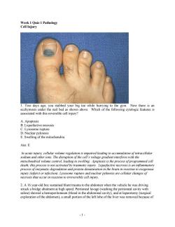
- 1 - Week 1 Quiz 1 Pathology Cell Injury 1. Two days ago, you stubbed your big toe while hurrying to the gym. Now there is an ecchymosis under the nail bed as shown above. Which of the following cytologic features is associated with this reversible cell injury? A. Apoptosis B. Liquefactive necrosis C. Lysosome rupture D. Nuclear pyknosis E. Swelling of the mitochondria Ans: E In acute injury, cellular volume regulation is impaired leading to accumulation of intracellular sodium and other ions. The disruption of the cell’s voltage gradient interferes with the mitochondrial volume control, leading to swelling. Apoptosis is the process of programmed cell death; this process is not activated by traumatic injury. Liquefactive necrosis is an inflammatory process of enzymatic degradation and protein denaturation in the brain in reaction to exogenous injury (infarct or infection). Lysosome rupture and nuclear pyknosis are cellular changes of necrosis that occur in reaction to irreversible cell injury. 2. A 16 year-old boy sustained blunt trauma to the abdomen when the vehicle he was driving struck a bridge abutment at high speed. Peritoneal lavage (washing the peritoneal cavity with saline) showed a hemoperitoneum (blood in the abdominal cavity), and at laparotomy (surgical exploration of the abdomen), a small portion of the left lobe of the liver was removed because of
the injury.Several weeks later,a CT scan of the abdomen shows that the liver is nearly normal in size again.Which of the following processes best explains this phenomenon? A.Apoptosis B.Dysplasia C.Fatty change D.Hydropic change E.Hyperplasia Ans:E in the mber of cells.which in this case is ar enter l grow py.Apoptosis is shape,and size.Fatty change (abnormal accumulation of lipids in parenchyma cells)usually occurs in the liver.In the heart,it would occur in the myocardium,not on the valves.Hydropic change is acute.reversible cell swelling in response to injury. 3.On a routine visit to the physician,an otherwise healthy 51 year-old man has an elevated blood pressure of 150/95 mm Hg.If the patient's hypertension remains untreated for years, which of the following cellular alterations will most likely be seen in the myocardium(heart muscle)? A.Atrophy B.Hemosiderosis C.Hypertrophy D.Metaplasia E Sclerosis Ans:C places increased stress on the lejt ventricle.which msi increase contractlejorce a pres. not possible. Instea cells in number or size of cells due to decreased stimulation or stress. an iron overload disorder unrelated to hypertension.Metaplasia is replacement ofone cell type with another due to irritation or environmental exposure.Sclerosis describes a hardening ofa structure,often due to replacement with connective tissue. -2
- 2 - the injury. Several weeks later, a CT scan of the abdomen shows that the liver is nearly normal in size again. Which of the following processes best explains this phenomenon? A. Apoptosis B. Dysplasia C. Fatty change D. Hydropic change E. Hyperplasia Ans: E Hyperplasia describes the increase in the number of cells, which in this case is a response to injury. It requires stimulation of cell sin G0 to enter the cell cycle and multiply. Apoptosis is programmed cell death. Dysplasia refers to abnormal growth of cells and loss of proper orientation, shape, and size. Fatty change (abnormal accumulation of lipids in parenchymal cells) usually occurs in the liver. In the heart, it would occur in the myocardium, not on the valves. Hydropic change is acute, reversible cell swelling in response to injury. 3. On a routine visit to the physician, an otherwise healthy 51 year-old man has an elevated blood pressure of 150/95 mm Hg. If the patient’s hypertension remains untreated for years, which of the following cellular alterations will most likely be seen in the myocardium (heart muscle)? A. Atrophy B. Hemosiderosis C. Hypertrophy D. Metaplasia E. Sclerosis Ans: C Hypertension places increased stress on the left ventricle, which must increase contractile force to eject blood against increased pressure. Cardiac tissue is terminally differentiated, thus hyperplasia is not possible. Instead, the cells increase in size (hypertrophy). Atrophy is a reduction in the number or size of cells due to decreased stimulation or stress. Hemosiderosis is an iron overload disorder unrelated to hypertension. Metaplasia is replacement of one cell type with another due to irritation or environmental exposure. Sclerosis describes a hardening of a structure, often due to replacement with connective tissue
©Elsevier2005 4.A 72 vear-old man died suddenly and unexpectedly from congestive heart failure.At autopsy the heart was very enlarged and weighed 580 grams.There was marked left ventricular hypertrophy and minimal oro arterial atherosclerosis.A serum chemistry panel ordered ath sho malities.Which of the followi ing the figure processes best accounts for the appearance of his aortic valve,which is seen Amyloidosis Dystrophic Fatty Ch ange D.Hemosiderosis E.Lipofuscin deposition Ans:B Dystrophic calcification is an extracellular deposition of calcium in the setting of persistent necrotic tissue.The figure shows calcium deposits on the aortic valve leaflets that impair blood flow,causing left ventricular hypertrophy.Amyloidosis is a large category of disorders characterized by diverse extracellular protein denosits not visible to the naked eve fatty chang (abnormal accumulation of lipids in parenchymal cells)usually occurs in the liver.In the heart, it would occur in the mvocardium,not on the valves.Hemosiderosis is an iron overload disorder that does not cause extracellular calcium deposition.Lipofuscin is the result of accumulated oxidized and cross-linked proteins:these depo sits are not visible to the naked eye. -3
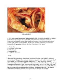
- 3 - 4. A 72 year-old man died suddenly and unexpectedly from congestive heart failure. At autopsy, the heart was very enlarged and weighed 580 grams. There was marked left ventricular hypertrophy and minimal coronary arterial atherosclerosis. A serum chemistry panel ordered prior to death showed no abnormalities. Which of the following pathologic processes best accounts for the appearance of his aortic valve, which is seen in the figure? A. Amyloidosis B. Dystrophic calcification C. Fatty Change D. Hemosiderosis E. Lipofuscin deposition Ans: B Dystrophic calcification is an extracellular deposition of calcium in the setting of persistent necrotic tissue. The figure shows calcium deposits on the aortic valve leaflets that impair blood flow, causing left ventricular hypertrophy. Amyloidosis is a large category of disorders characterized by diverse extracellular protein deposits not visible to the naked eye. Fatty change (abnormal accumulation of lipids in parenchymal cells) usually occurs in the liver. In the heart, it would occur in the myocardium, not on the valves. Hemosiderosis is an iron overload disorder that does not cause extracellular calcium deposition. Lipofuscin is the result of accumulated oxidized and cross-linked proteins; these deposits are not visible to the naked eye
©Elsevier2005 5.A38 year-old woman experienced severe abdominal pain with hypotension and shock that led to her death within 36 hours after the onset of the pain.An autopsy was performed.The mesentery is shown above.The fatty tissue is covered with multiple yellow areas of tissue injury. Which of the following events has most likely occurred? A.Acute pancreatitis B.Gangrenous cholecystitis C.Hepatitis B virus infection D.Small intestinal infarction E.Tuberculous lymphadenitis Ans:A e pancreatitis which causes en chalk matic fat ne the would sh n by ecro organ ns. clinical features would have more green-bioo ikely b RUO pain an orexia,but no no association of pancreatitis and Hepatitis B infection.although case reports have suggested an increased mortality in the event of co-infection.Small bowel infarction would demonstrate dusky serosa and hemorrhage,not chalky-white nodules.Lymphadenitis caused by tuberculosis is most commonly of the cervical nodes,but infection of peritoneal,peri-pancreatic,mesenteric,or hepatic lymph nodes is possible.These infections are unlikely to lead to rapidly progressive shock and death.For example,hepatic nodal involvement might cause slow onset of jaundice and portal hypertension. -4
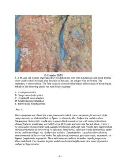
- 4 - 5. A 38 year-old woman experienced severe abdominal pain with hypotension and shock that led to her death within 36 hours after the onset of the pain. An autopsy was performed. The mesentery is shown above. The fatty tissue is covered with multiple yellow areas of tissue injury. Which of the following events has most likely occurred? A. Acute pancreatitis B. Gangrenous cholecystitis C. Hepatitis B virus infection D. Small intestinal infarction E. Tuberculous lymphadenitis Ans: A These symptoms are classic for acute pancreatitis which causes enzymatic fat necrosis of the peri-pancreatic or abdominal fat via lipase, as shown by the chalky-white nodules above. Gangrenous cholecystitis would show a green-black necrotic organ with some perforations; clinical features would have more likely been RUQ pain and anorexia, but not shock. There is no association of pancreatitis and Hepatitis B infection, although case reports have suggested an increased mortality in the event of co-infection. Small bowel infarction would demonstrate dusky serosa and hemorrhage, not chalky-white nodules. Lymphadenitis caused by tuberculosis is most commonly of the cervical nodes, but infection of peritoneal, peri-pancreatic, mesenteric, or hepatic lymph nodes is possible. These infections are unlikely to lead to rapidly progressive shock and death. For example, hepatic nodal involvement might cause slow onset of jaundice and portal hypertension
6.In an experiment,cells are subjected to radiant energy in the form of x-rays This results in cell injury caused by hydrolysis of water and free radical generation.Which of the following cellular enzymes normally protects cells from free radical injury? A.Endonuclease B.Glutathione peroxidase C.Lactate dehydrogenase D.Phosholipase E.Protease Ans:B Glutathione peroxidase is one of three key enzymes that neutralize free radicals (the others are superoxide dismuase and caalase).Endonuclease cleaves phosphodiester bonds chain of tides. Lactate dehydrogend 2 the re nate to e and the fatty acids. e bonds in the process ofproteolysis robzes lipids 7.A30 year old man fract tures his left femur in a biking a cide d his leg is pl ced in a plaster cast. fter the leg has been immobilized for several week the diameter the left cal This change is most likely to result from which of the following alterations in the calf muscles A.Aplasia B.Atrophy C.Dystrophy D.Hypoplasia E.Metaplasia Ans:B Atrophy refers to the reduction in n umber or size ofcells.In this case,disuse of the limb results d neuronal stimulation to the skeletal n Aplasia is the failr of his leg. which leads to atre etof元 m D the dege of tissue Hypop acement ofone cell type with another in response to stress or irritation 8. A experiment ana yme activity associated wit A.Endothelial cells B.Erythrocytes C.Germ cells D.Neurons E.Neutrophils 5
- 5 - 6. In an experiment, cells are subjected to radiant energy in the form of x-rays. This results in cell injury caused by hydrolysis of water and free radical generation. Which of the following cellular enzymes normally protects cells from free radical injury? A. Endonuclease B. Glutathione peroxidase C. Lactate dehydrogenase D. Phosholipase E. Protease Ans: B Glutathione peroxidase is one of three key enzymes that neutralize free radicals (the others are superoxide dismutase and catalase). Endonuclease cleaves phosphodiester bonds of a chain of nucleotides. Lactate dehydrogenase catalyzes the reduction of pyruvate to lactate and the oxidation of lactate to pyruvate in the liver (Cori cycle). Phospholipase hydrolyzes lipids into fatty acids. Protease hydrolyzes peptide bonds in the process of proteolysis. 7. A 30 year-old man fractures his left femur in a biking accident, and his leg is placed in a plaster cast. After the leg has been immobilized for several weeks, the diameter of the left calf has decreased. This change is most likely to result from which of the following alterations in the calf muscles? A. Aplasia B. Atrophy C. Dystrophy D. Hypoplasia E. Metaplasia Ans: B Atrophy refers to the reduction in number or size of cells. In this case, disuse of the limb results in decreased neuronal stimulation to the skeletal muscles of his leg, which leads to atrophy. Aplasia is the failure of a structure to form. Dystrophy is the degeneration of tissue due to a pathologic process. Hypoplasia refers to incomplete development of a tissue or organ. Metaplasia is replacement of one cell type with another in response to stress or irritation. 8. An experiment analyzes cells for enzyme activity associated with sustained cellular proliferation. Which of the following cell types is most likely to have the highest telomerase activity? A. Endothelial cells B. Erythrocytes C. Germ cells D. Neurons E. Neutrophils
Ans:C Telomerase is a eukaryotic reverse transcriptase that adds nucleotide repeats to the 3'end of DNA.Chromosomes would otherwise be progressively shortened due to inability of DNA polymerase to replication without a primer.Germ cells express telomerase but permanent cells do not.Endothelial cells.neurons,and neutrophils are terminally differentiated cells and do not express telomerase.Erythrocytes lack a nucleus and therefore telomerase has no function. ©Elsevier2005 9.A 52 year-old man frequently experiences"heartburn"and gastric reflux after eating After tin copy A biopsy of the ogic changes,seen in A.Atrophy of lamina propria B.Gastric epithelial metaplasia C.Goblet cell hyperplasia D.Mucosal hypertrophy E.Squamous transformation Ans:B The esophagus is lined by stratified squamous epithelial,but in the presence ofpersistent irritation s ch as gastric reflux.metar olasia occurs which replaces the squamor epithelia with pithelia typical of the gastric mucosa.The lamind al in size with respect to the colu r cells.The encompasses the epi thelial layer lamina propria. and muscularis mucosa:the latter -6
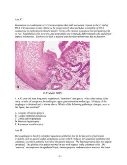
- 6 - Ans: C Telomerase is a eukaryotic reverse transcriptase that adds nucleotide repeats to the 3’ end of DNA. Chromosomes would otherwise be progressively shortened due to inability of DNA polymerase to replication without a primer. Germ cells express telomerase but permanent cells do not. Endothelial cells, neurons, and neutrophils are terminally differentiated cells and do not express telomerase. Erythrocytes lack a nucleus and therefore telomerase has no function. 9. A 52 year-old man frequently experiences “heartburn” and gastric reflux after eating. After many mouths of symptoms, he undergoes upper gastrointestinal endoscopy. A biopsy of the esophagus is obtained and is shown above. Which of the following pathologic changes, seen in the figure, has occurred?’ A. Atrophy of lamina propria B. Gastric epithelial metaplasia C. Goblet cell hyperplasia D. Mucosal hypertrophy E. Squamous transformation Ans: B The esophagus is lined by stratified squamous epithelial, but in the presence of persistent irritation such as gastric reflux, metaplasia occurs which replaces the squamous epithelia with columnar, secretory epithelia typical of the gastric mucosa. The lamina propria does not appear atrophied. The globlet cells appear normal in size with respect to the columnar cells. The “mucosa” encompasses the epithelial layer, lamina propria, and muscularis mucosa; the latter
wo layers are not hypertrophied."Transfo matio "usually refers to the transformation one f the cervix where stratified squamous epithelial of the ectocervix meets simple columnar epithelia of the endocervix.In the context of the GI tract,"squamous transformation"is a tempting choice, but it is an imprecise term referring to metaplasia. 10.On day 28 of her menstrual cycle,a 23 year-old woman experiences onset of menstrual bleeding that lasts for 6 days.She has had regular cycles for many years.Which of the following processes is most likely occurring in the endometrium just before the onset of normal menstrual bleeding? A.Apoptosis B.Atrophy C Caseous necrosis D.Heterophagocytosis E.Liquef active necros Ans:A is a physiologic process that occurs in the endometrium in absence ofpregnancy-induced ho ation.During menstruation,layers of the ometrium are shed:they do not shrink in size.Caseous necrosis is a type of pathologic cel death characteristic of fungal or Mycobacterium tuberculosis infections.Heterphagocytosis is the process by which neutrophils and macrophages digest bacteria and apoptotic bodies. Liquefactive necrosis is an inflammatory process of enzymatic degradation and protein denaturation in the brain in reaction to exogenous injury (infarct or infection). 11.A 33 year-old woman was admitted to the hospital for a hysterectomy.During surgery she experienced an episode of profound hypotension.She was apparently improving after this episode.Now she has increasing letharg y and decreased urine output.Laborato ry studies to evaluate renal function are abnd rmal and show an elevated serum creatinine level of 4.3 mg/dL and elevated urea nitrogen level of 40 mg/dL.A renal biopsy is perfo xamined using electron micro py.Which of the follow diagnosis of acute tubular necro osis? Chr drial D.Pla sma m rane ble Ans:C Nuclear fragmentation is an indication of irreversible cell injury that leads to necrosis. Chromatin clumping,Mitochondrial swelling,Plasma membrane blebs and Ribosomal disaggregation are common to both reversible and irreversible cell injury.Reversible injury would not lead to necrosis. .7
- 7 - two layers are not hypertrophied. “Transformation” usually refers to the transformation zone of the cervix where stratified squamous epithelial of the ectocervix meets simple columnar epithelia of the endocervix. In the context of the GI tract, “squamous transformation” is a tempting choice, but it is an imprecise term referring to metaplasia. 10. On day 28 of her menstrual cycle, a 23 year-old woman experiences onset of menstrual bleeding that lasts for 6 days. She has had regular cycles for many years. Which of the following processes is most likely occurring in the endometrium just before the onset of normal menstrual bleeding? A. Apoptosis B. Atrophy C. Caseous necrosis D. Heterophagocytosis E. Liquefactive necrosis Ans: A Apoptosis, programmed cell death, is a physiologic process that occurs in the endometrium in the absence of pregnancy-induced hormonal stimulation. During menstruation, layers of the endometrium are shed; they do not shrink in size. Caseous necrosis is a type of pathologic cell death characteristic of fungal or Mycobacterium tuberculosis infections. Heterphagocytosis is the process by which neutrophils and macrophages digest bacteria and apoptotic bodies. Liquefactive necrosis is an inflammatory process of enzymatic degradation and protein denaturation in the brain in reaction to exogenous injury (infarct or infection). 11. A 33 year-old woman was admitted to the hospital for a hysterectomy. During surgery she experienced an episode of profound hypotension. She was apparently improving after this episode. Now she has increasing lethargy and decreased urine output. Laboratory studies to evaluate renal function are abnormal and show an elevated serum creatinine level of 4.3 mg/dL and elevated urea nitrogen level of 40 mg/dL. A renal biopsy is performed, and the specimen is examined using electron microscopy. Which of the following morphologic changes most likely suggests a diagnosis of acute tubular necrosis? A. Chromatin clumping B. Mitochondrial swelling C. Nuclear fragmentation D. Plasma membrane blebs E. Ribosomal disaggregation Ans: C Nuclear fragmentation is an indication of irreversible cell injury that leads to necrosis. Chromatin clumping, Mitochondrial swelling, Plasma membrane blebs and Ribosomal disaggregation are common to both reversible and irreversible cell injury. Reversible injury would not lead to necrosis
12.Which of the following statements is correct? A.Autophagy is a rare process that is always associated with disease. B.Autophagy is important in cellular defense against microorganisms. C.Autophagy is not involved in cellular differentiation. D.The autophagic vacuole fuses with a lysosome to form an autophagolysosome E.The autophagic vacuole is formed by mitochondria. Ans:D After a pha osome full ofproteolytic b organelle acrophages engul 90pagRaaC9mpicaei3gnorraeOaemror8am on of RBCs,B-cells,adipocytes,osteoclasts,and keratinocytes.This process is critical to cellular remodeling.The vacuole results from endocytosis at the plasma membrane,not the mitochrondria. -8
- 8 - 12. Which of the following statements is correct? A. Autophagy is a rare process that is always associated with disease. B. Autophagy is important in cellular defense against microorganisms. C. Autophagy is not involved in cellular differentiation. D. The autophagic vacuole fuses with a lysosome to form an autophagolysosome. E. The autophagic vacuole is formed by mitochondria. Ans: D After a phagosome has been internalized into the cell, it fuses with a lysosome full of proteolytic enzymes that will degrade the contents of the vacuole. Autophagy is a physiologic process by which a cell digests its own unnecessary or dysfunctional organelles. Macrophages engulf bacteria via phagocytosis, not autophagy, in order to destroy invasive microorganisms. Autophagy has been implicated differentiation of RBCs, B-cells, adipocytes, osteoclasts, and keratinocytes. This process is critical to cellular remodeling. The vacuole results from endocytosis at the plasma membrane, not the mitochrondria
按次数下载不扣除下载券;
注册用户24小时内重复下载只扣除一次;
顺序:VIP每日次数-->可用次数-->下载券;
- 同济大学:《眼科学》课程电子教案(PPT课件讲稿)眼肿瘤 Ocular trauma.ppt
- 同济大学:《眼科学》课程电子教案(PPT课件讲稿)青光眼 Glaucoma.pptx
- 同济大学:《眼科学》课程电子教案(PPT课件讲稿)角膜病总论 Corneal disease.ppt
- 同济大学:《眼科学》课程电子教案(PPT课件讲稿)视觉系统 Visual Organ.ppt
- 同济大学:《眼科学》课程电子教案(PPT课件讲稿)视网膜疾病 Retinal Disease.ppt
- 同济大学:《眼科学》课程电子教案(PPT课件讲稿)视光学基础 Basic Optics.ppt
- 同济大学:《眼科学》课程电子教案(PPT课件讲稿)葡萄膜炎 Disease of the Uvea.ppt
- 同济大学:《眼科学》课程电子教案(PPT课件讲稿)眼组织学 Histology of the Eye.ppt
- 同济大学:《眼科学》课程电子教案(PPT课件讲稿)眼科导论 Introduction(负责人:徐国彤).ppt
- 同济大学:《眼科学》课程教学资源(大纲教案)眼科学教学大纲(英文,Ophthalmology).doc
- 同济大学:《眼科学》课程教学资源(大纲教案)眼科学教学大纲(中文,负责人:徐国彤).doc
- 同济大学:《眼科学》课程电子教案(PPT课件讲稿)白内障 Ophthalmology.ppt
- 同济大学:《眼科学》课程电子教案(PPT课件讲稿)眼睑和眼表疾病 The eyelids & ocular surface diseases.ppt
- 同济大学:《眼科学》课程电子教案(PPT课件讲稿)眼眶异常 Disorders of the orbit.ppt
- 同济大学:《眼科学》课程电子教案(PPT课件讲稿)斜视 STRABISMUS.ppt
- 同济大学:《眼科学》课程电子教案(PPT课件讲稿)巩膜病 Scleral Disease.ppt
- 同济大学:《眼科学》课程电子教案(PPT课件讲稿)屈光学 Optics and Refractive errors correction.ppt
- 同济大学:《眼科学》课程电子教案(PPT课件讲稿)屈光不正 Our eye as a camera Refraction, errors and solutions..ppt
- 同济大学:《眼科学》课程电子教案(PPT课件讲稿)泪道疾病 THE WATERING EYE.ppt
- 同济大学:《眼科学》课程教学资源(试卷习题)MBBS试题.doc
- 同济大学:《病理学》课程教学资源(试卷习题,含答案)Inflammation.pdf
- 同济大学:《病理学》课程教学资源(试卷习题,含答案)Neoplasia.pdf
- 同济大学:《病理学》课程教学资源(试卷习题,含答案)Thrombosis I-II, Hemodynamics, Atherosclerosis.pdf
- 同济大学:《病理学》课程教学资源(试卷习题,含答案)Cardiac pathology.pdf
- 同济大学:《病理学》课程教学资源(试卷习题,含答案)Female reproductivepathology.pdf
- 同济大学:《病理学》课程教学资源(试卷习题,含答案)GIpathology.pdf
- 同济大学:《病理学》课程教学资源(试卷习题,含答案)试题A卷.pdf
- 同济大学:《病理学》课程教学资源(试卷习题,含答案)试题B卷.pdf
- 同济大学:《病理学》课程教学资源(教案讲义)Introduction to Pathology.pdf
- 同济大学:《病理学》课程教学资源(教案讲义)Chapter 01 Adaptation and Injury of Cell and Tissue(Adaptation of Cell and Tissue).pdf
- 同济大学:《病理学》课程教学资源(教案讲义)Chapter 01 Adaptation and Injury of Cell and Tissue(Reversible Injury of Cell and Tissue).pdf
- 同济大学:《病理学》课程教学资源(教案讲义)Chapter 01 Adaptation and Injury of Cell and Tissue(Irreversible Injury of Cell and Tissue).pdf
- 同济大学:《病理学》课程教学资源(教案讲义)Chapter 02 组织再生与修复 Tissue regeneration and repair.pdf
- 同济大学:《病理学》课程教学资源(教案讲义)Chapter 14 女性生殖系统和乳房疾病 THE DISEASE OF FEMALE GENITAL SYSTEM AND BREAST.pdf
- 同济大学:《病理学》课程教学资源(教案讲义)Chapter 16 神经系统疾病 Diseases of the Nervous System.pdf
- 吉林大学:《医学微生物学》课程电子教案(PPT课件)第一章 绪论、第一篇 细菌学总论 第一章 细菌的形态学(负责人:侯芳玉).ppt
- 吉林大学:《医学微生物学》课程电子教案(PPT课件)第二章 细菌的生理.ppt
- 吉林大学:《医学微生物学》课程电子教案(PPT课件)第三章 消毒与灭菌.ppt
- 吉林大学:《医学微生物学》课程电子教案(PPT课件)第五章 细菌的遗传与变异.ppt
- 吉林大学:《医学微生物学》课程电子教案(PPT课件)第六章 细菌的感染与致病机制(6.1-6.3).ppt
