南方医科大学:《内科学》课程教学课件(讲稿)第二篇 呼吸系统疾病 第十二章 胸膜疾病 Diseases of the Pleura
DISEASES OF PLEURA Pleural effusion 南方医科大学江医院 胸腔积液 呼吸科 pneumothorax 夏虎 气胸
Pleural effusion 胸腔积液 pneumothorax 气胸 南方医科大学江医院 呼吸科 夏虎 DISEASES OF PLEURA
PLEURA The pleura,which is a thin membrane that covers the inner surfaces of the thoracic cavity(胸腔),consists of a layer of mesothelial cells(间皮细胞)supported by a network of connective and fibroelastic tissue.The visceral pleura (脏层胸膜)lines the lung,.whereas the parietal pleura(壁 层胸膜)lines the rib cage,diaphragm,and mediastinal structures. The closed space between the visceral and parietal pleura is referred to as the pleural space.The vascular supply of the parietal pleural surface is from the systemic circulation(体循环),and it contains sensory nerves and lymphatics.By contrast,the visceral pleura is supplied with blood vessels from the pulmonary circulation(肺循环) and has no sensory nerves
第 一 部 分 The pleura, which is a thin membrane that covers the inner surfaces of the thoracic cavity(胸腔), consists of a layer of mesothelial cells(间皮细胞) supported by a network of connective and fibroelastic tissue. The visceral pleura (脏层胸膜)lines the lung,whereas the parietal pleura(壁 层胸膜) lines the rib cage, diaphragm, and mediastinal structures. The closed space between the visceral and parietal pleura is referred to as the pleural space. The vascular supply of the parietal pleural surface is from the systemic circulation(体循环), and it contains sensory nerves and lymphatics. By contrast, the visceral pleura is supplied with blood vessels from the pulmonary circulation(肺循环) and has no sensory nerves. PLEURA
PLEURAL EFFUSION EPIDEMIOLOGY AND PATHOBIOLOGY Pleural effusions occur most frequently in patients with pneumonia(肺炎)or heart failure(心衰). Normally,a small amount of fluid in the pleural space forms a thin layer between the visceral and parietal pleural surfaces and acts as a lubricant(润滑剂)to minimize friction between the chest wall and lung as they move against each other during inspiration (and expiration (呼气). This flux of fluid depends on the oncotic pressure(渗透压) and hydrostatic pressure(静水压)_within the parietal and visceral pleura as well as the pressure within the pleural space itself
PLE第URA一L E部FF分USION EPIDEMIOLOGY AND PATHOBIOLOGY Pleural effusions occur most frequently in patients with pneumonia(肺炎) or heart failure(心衰). Normally, a small amount of fluid in the pleural space forms a thin layer between the visceral and parietal pleural surfaces and acts as a lubricant(润滑剂) to minimize friction between the chest wall and lung as they move against each other during inspiration(吸气) and expiration (呼气). This flux of fluid depends on the oncotic pressure (渗透压) and hydrostatic pressure (静水压) within the parietal and visceral pleura as well as the pressure within the pleural space itself
PLEURAL EFFUSION Hydrostatic pressure in the parietal pleura is similar to systemic circulation(30 cm H20),whereas that of the visceral pleura is similar to the pulmonary circulation(10 cm H20).Accordingly,most fluid in the pleural space is filtered from parietal pleura.Because the pressure within the pleural space itself is more subatmospheric at the apex than at the base,most of the fluid filters in from the less dependent upper lung zones. Fluid is drained out primarily through lymphatics in the parietal pleura.The fluid enters through lymphatic stomas (淋巴孔)on the surface of the parietal pleura,which are located beneath the mesothelial monolayer.The normal turnover of fluid within the pleural space is 10 to 20 mL/day
第 一 部 分 Hydrostatic pressure in the parietal pleura is similar to systemic circulation (30 cm H2O), whereas that of the visceral pleura is similar to the pulmonary circulation (10 cm H2O). Accordingly, most fluid in the pleural space is filtered from parietal pleura. Because the pressure within the pleural space itself is more subatmospheric at the apex than at the base, most of the fluid filters in from the less dependent upper lung zones. Fluid is drained out primarily through lymphatics in the parietal pleura. The fluid enters through lymphatic stomas (淋巴孔)on the surface of the parietal pleura, which are located beneath the mesothelial monolayer. The normal turnover of fluid within the pleural space is 10 to 20 mL/day. PLEURAL EFFUSION
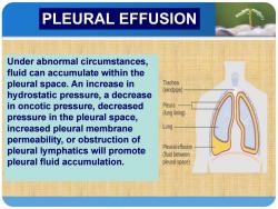
PLEURAL EFFUSION Under abnormal circumstances, fluid can accumulate within the pleural space.An increase in Trachea (windpipe) hydrostatic pressure,a decrease in oncotic pressure,decreased Pleura (lung lining) pressure in the pleural space, increased pleural membrane Lung permeability,or obstruction of pleural lymphatics will promote Pleural effusion (fluid between pleural fluid accumulation. pleural space)
第 一 部 分 Under abnormal circumstances, fluid can accumulate within the pleural space. An increase in hydrostatic pressure, a decrease in oncotic pressure, decreased pressure in the pleural space, increased pleural membrane permeability, or obstruction of pleural lymphatics will promote pleural fluid accumulation. PLEURAL EFFUSION
PLEURAL EFFUSION An increase in hydrostatic pressure or decrease in oncotic pressure will result in a low-protein collection of pleural fluid characterized as transudates(漏出液).For example,heart failure can produce transudates by increasing hydrostatic pressure in the pulmonary venous system,atelectasis(肺不张)_can promote transudates by making pleural pressure more subatmospheric,and occasionally, oncotic pressure may be suffciently reduced to cause transudates (hypoalbuminemia低白蛋白血症). Changes in pleural membrane permeability can produce high-protein effusions,which are characterized as exudates(渗出液)and can be seen in inflammatory states such as pneumonia,tuberculosis 痼),or rheumatoid arthritis(类风湿关节炎) Tumors can disrupt the integrity of the mesothelial layer or the integrity of the capillary epithelium,thereby resulting in exudative effusions,or they may block lymphatic drainage
第 一 部 分 An increase in hydrostatic pressure or decrease in oncotic pressure will result in a low-protein collection of pleural fluid characterized as transudates(漏出液). For example, heart failure can produce transudates by increasing hydrostatic pressure in the pulmonary venous system, atelectasis(肺不张) can promote transudates by making pleural pressure more subatmospheric, and occasionally, oncotic pressure may be suffciently reduced to cause transudates (hypoalbuminemia低白蛋白血症). Changes in pleural membrane permeability can produce high-protein effusions, which are characterized as exudates(渗出液) and can be seen in inflammatory states such as pneumonia, tuberculosis(结核 病), or rheumatoid arthritis(类风湿关节炎). Tumors can disrupt the integrity of the mesothelial layer or the integrity of the capillary epithelium, thereby resulting in exudative effusions, or they may block lymphatic drainage. PLEURAL EFFUSION

PLEURAL EFFUSION CLINICAL MANIFESTATIONS Patients with pleural effusions may be asymptomatic 状)or may experience dyspnea(呼吸困难).When the parietal pleura is actively inflamed,pain can be present, and it is generally unilateral(单侧),sharp,and worsens with inspiration. At times,effusions may be suffciently large to contribute to respiratory failure(呼吸衰竭).Physical findings include dullness to percussion(叩诊浊音)in the area of the effusion,along with diminished breath sounds(呼吸音减弱) and absent tactile fremitus(触觉语颤)
第 一 部 分 CLINICAL MANIFESTATIONS Patients with pleural effusions may be asymptomatic(无症 状) or may experience dyspnea(呼吸困难). When the parietal pleura is actively inflamed, pain can be present, and it is generally unilateral(单侧), sharp, and worsens with inspiration. At times, effusions may be suffciently large to contribute to respiratory failure(呼吸衰竭). Physical findings include dullness to percussion (叩诊浊音)in the area of the effusion, along with diminished breath sounds(呼吸音减弱) and absent tactile fremitus(触觉语颤). PLEURAL EFFUSION
PLEURAL EFFUSION DIAGNOSIS Chest radiography is often the first imaging method used to detect an effusion.The volume of fluid in the pleural space needs to exceed 250 mL to be visualized on the chest radiograph.When an effusion is present,there is blunting of the costophrenic angle(肋膈角)on the posteroanterior chest radiograph,and a meniscus(新月液面) can be seen posteriorly on the lateral chest radiograph. Fluid may also collect in either the minor or major fissures
第 一 部 分 DIAGNOSIS Chest radiography is often the first imaging method used to detect an effusion. The volume of fluid in the pleural space needs to exceed 250 mL to be visualized on the chest radiograph. When an effusion is present, there is blunting of the costophrenic angle(肋膈角) on the posteroanterior chest radiograph, and a meniscus(新月液面) can be seen posteriorly on the lateral chest radiograph. Fluid may also collect in either the minor or major fissures. PLEURAL EFFUSION

PLEURAL EFFUSION Occasionally,pleural fluid collections in the major or minor fissures may appear as a pulmonary mass(肺部团块) and are referred to as pseudotumors ()A lateral Decubitus(侧卧位)chest radiograph can be obtained to determine whether fluid is free flowing or loculated 的). Chest CT provides better characterization of pleural and parenchymal abnormalities by better defining loculated effusions,distinguishing between atelectasis (and effusion,and distinguishing loculated effusion from lung abscess(肺脓肿)
第 一 部 分 Occasionally, pleural fluid collections in the major or minor fissures may appear as a pulmonary mass(肺部团块) and are referred to as pseudotumors(假瘤). A lateral Decubitus(侧卧位) chest radiograph can be obtained to determine whether fluid is free flowing or loculated(包裹 的). Chest CT provides better characterization of pleural and parenchymal abnormalities by better defining loculated effusions, distinguishing between atelectasis(不张) and effusion, and distinguishing loculated effusion from lung abscess(肺脓肿) PLEURAL EFFUSION

PLEURAL EFFUSION 101920 CHEST PAW 1g/19/209994982 95 UPRIGHT
PLE第URA一L E部FF分USION
按次数下载不扣除下载券;
注册用户24小时内重复下载只扣除一次;
顺序:VIP每日次数-->可用次数-->下载券;
- 南方医科大学:《内科学》课程教学课件(讲稿)第二篇 呼吸系统疾病 第九章 间质性肺病(ILD).pdf
- 南方医科大学:《内科学》课程教学课件(讲稿)第二篇 呼吸系统疾病 第三章 慢性支气管炎——慢性阻塞性肺疾病 Chronic Obstructive Pulmonary Disease(英文版).pdf
- 南方医科大学:《内科学》课程教学课件(讲稿)第二篇 呼吸系统疾病 第三章 慢性阻塞性肺疾病(Chronic obstructive pulmonary disease,COPD).pdf
- 南方医科大学:《内科学》课程教学课件(讲稿)第二篇 呼吸系统疾病 第七章 肺结核 Pulmonary tuberculosis(主讲:龚雨新).pdf
- 南方医科大学:《内科学》课程教学课件(讲稿)第一篇 内科学绪论 Introduction to Internal Medicine.pdf
- 《内科学》课程教学资源(新冠文献)The global spread of 2019-nCoV - a molecular evolutionary analysis.pdf
- 《内科学》课程教学资源(新冠文献)Potent binding of 2019 novel coronavirus spike protein by a SARS coronavirus-specific human monoclonal antibody.pdf
- 《内科学》课程教学资源(新冠文献)Molecular and serological investigation of 2019- nCoV infected patients - implication of multiple shedding routes.pdf
- 《内科学》课程教学资源(新冠文献)HIV-1 did not contribute to the 2019-nCoV genome.pdf
- 《内科学》课程教学资源(新冠文献)Emerging novel coronavirus(2019-nCoV)current scenario, evolutionary perspective based on genome analysis and recent developments.pdf
- 《内科学》课程教学资源(新冠文献)Early Transmission Dynamics in Wuhan, China, of Novel Coronavirus–Infected Pneumonia.pdf
- 《内科学》课程教学资源(新冠文献)Clinical features of patients infected with 2019 novel coronavirus in Wuhan, China.pdf
- 《内科学》课程教学资源(新冠文献)Clinical Characteristics of Coronavirus Disease 2019 in China.pdf
- 《内科学》课程教学资源(新冠文献)An emerging coronavirus causing pneumonia outbreak in Wuhan, China - calling for developing therapeutic and prophylactic strategies.pdf
- 《内科学》课程教学资源(新冠文献)A Novel Coronavirus from Patients with Pneumonia in China, 2019.pdf
- 南方医科大学:《内科学》课程教学课件(PPT讲稿)第二篇 呼吸系统疾病 第十章 急性肺栓塞(肺血栓栓塞症 Pulmonary thromboembolism).pptx
- 南方医科大学:《内科学》课程教学课件(PPT讲稿)第二篇 呼吸系统疾病 第九章 间质性肺疾病 Interstitial Lung Diseases(主讲:张海云).pptx
- 南方医科大学:《内科学》课程教学课件(PPT讲稿)第二篇 呼吸系统疾病 第八章 肺癌 Lung cancer(主讲:吴锡平).pptx
- 南方医科大学:《内科学》课程教学大纲 Internal medicine(共八篇,临床医学五年制本科专业).doc
- 山东第一医科大学(泰山医学院):《优生优育》课程作业习题(打印版)第十二章 儿童心理行为异常(含答案).pdf
- 南方医科大学:《内科学》课程教学课件(讲稿)第二篇 呼吸系统疾病 第十章 肺血栓栓塞症(主讲:阳倩捷).pdf
- 南方医科大学:《内科学》课程教学课件(讲稿)第二篇 呼吸系统疾病 第十五章 呼吸衰竭与ARDS(呼吸衰竭与呼吸支持技术以及急性呼吸窘迫综合征).pdf
- 人民卫生出版社:《内科学》课程电子教材(循环系统疾病)第七章 先心性心血管病.pdf
- 人民卫生出版社:《内科学》课程电子教材(循环系统疾病)第四章 动脉粥样硬化和冠状动脉粥样硬化性心脏病.pdf
- 人民卫生出版社:《内科学》课程电子教材(循环系统疾病)第三章 心律失常.pdf
- 人民卫生出版社:《内科学》课程电子教材(循环系统疾病)第六章 心肌疾病.pdf
- 人民卫生出版社:《内科学》课程电子教材(循环系统疾病)第八章 心脏瓣膜病.pdf
- 人民卫生出版社:《内科学》课程电子教材(循环系统疾病)第一章 心血管总论、第二章 心力衰竭.pdf
- 人民卫生出版社:《内科学》课程电子教材(循环系统疾病)第五章 高血压.pdf
- 南方医科大学:《内科学》课程教学课件(讲稿)第三篇 循环系统疾病 第四章 冠状动脉粥样硬化性心脏病——动脉粥样硬化和冠心病(主讲:陈安).pdf
- 南方医科大学:《内科学》课程教学课件(讲稿)第三篇 循环系统疾病 第五章 高血压 Hypertention(主讲:缪绯).pdf
- 南方医科大学:《内科学》课程教学课件(讲稿)第三篇 循环系统疾病 第六章 心肌疾病(主讲:刘芃).pdf
- 南方医科大学:《内科学》课程教学课件(讲稿)第三篇 循环系统疾病 第八章 心脏瓣膜病 Valvular Heart Disease(主讲:张培东).pdf
- 南方医科大学:《内科学》课程教学课件(讲稿)第三篇 循环系统疾病 第九章 心包炎 Pericarditis.pdf
- 南方医科大学:《内科学》课程教学课件(讲稿)第三篇 循环系统疾病 第十章 感染性心内膜炎(Infective Endocarditis, IE).pdf
- 南方医科大学:《内科学》课程教学课件(讲稿)第四篇 消化系统疾病 第五章 消化性溃疡(Peptic Ulcer, PU).pdf
- 南方医科大学:《内科学》课程教学课件(讲稿)第四篇 消化系统疾病 第五章 消化性溃疡 peptic ulcer(主讲:唐银丽).pdf
- 南方医科大学:《内科学》课程教学课件(讲稿)第四篇 消化系统疾病 第六章 胃癌 Gastric Cancer(主讲:毛华).pdf
- 南方医科大学:《内科学》课程教学课件(讲稿)第四篇 消化系统疾病 第八章 炎症性肠病(主讲:王新颖).pdf
- 南方医科大学:《内科学》课程教学课件(讲稿)第四篇 消化系统疾病 第十五章 肝硬化 Cirrhosis of Liver(主讲:卢敏).pdf
