广东海洋大学:《兽医内科学》课程教学课件(讲稿)Chapter 4 Diseases of cardiovascular system

VIMChapter 4 Diseases of cardiovascular system$ChapterMainContentsPulmonarycapillarynetworkBlood CirculatorySystemBloodflowRightSystemiccaplllanynetworkGuangdong Ocean University
1 Chapter 4 Diseases of cardiovascular system Chapter Main Contents • Conspectus • Section 1 Acute heart failure • Section 2 Traumatic pericarditis
Chapter4Diseases of cardiovascularsystemVIM$Conspectus> The cardiovascular system comprises the heart, the veins, and thearteries.The cardiovascular system maintains normal systemicarterial pressure,bloodflowtometabolizing tissue,and capillary(systemicandpulmonary)pressures.The rate andforce of contraction of the heart and the degree of constriction ordilatation of blood vessels aredetermined by theautonomic nervous system and hormones produced either at theheart and blood vessels.Slightly >10% of all domestic animals examined by a veterinarianhave someform of cardiovascular disease.In addition, cardiovasculardiseases may be more difficult to detect and quantify because the heart cannot beseen and is protected so well by the rib cage.>Therefore,evaluationof theheartdepends onheartsoundsandmurmurs, pressure pulses and the apex beat, the electrocardiogram,and so on.Guangdong Ocean University
2 Conspectus Ø The cardiovascular system the heart, the veins, and the arteries. The cardiovascular system maintains normal systemic arterial pressure, blood flow to metabolizing tissue, and capillary (systemic and pulmonary) pressures. Ø The rate and force of contraction of the heart and the degree of constriction or dilatation of blood vessels are determined by the autonomic nervous system and hormones produced either at the heart and blood vessels. of all domestic animals examined by a veterinarian have some form of cardiovascular disease. In addition, cardiovascular diseases may be more difficult to because the heart cannot be seen and is protected so well by the rib cage. Ø Therefore, of the heart depends on heart sounds and murmurs, pressure pulses and the apex beat, the electrocardiogram, and so on
Chapter 4Diseases of cardiovascular systemVIMSection1AcuteHeartFailureOverviewHeart failure is not a specific disease or diagnosis,but asyndrome in which severe systolic and/or diastolic dysfunctionresults in decompensation of the cardiovascularsystem.Thereare limited and specific mechanisms by which heart disease can result indecompensation of thecardiovascular system.As aresult, there are limited andspecific clinical signs that can develop as a result of heart failure.> Heart failurecan be divided into 4functional classificationssystolic收缩的myocardial failure,impedance阻抗to cardiac inflow,pressure overload, and volume overload.To be continuedGuangdong Ocean University
3 Section 1 Acute Heart Failure uOverview is not a specific disease or diagnosis, but a syndrome in which severe systolic and/or diastolic dysfunction results in decompensation of the cardiovascular system. There are limited and specific mechanisms by which heart disease can result in decompensation of the cardiovascular system. As a result, there are limited and specific clinical signs that can develop as a result of heart failure. Ø Heart failure can be divided into 4 functional classifications: systolic收缩的 myocardial failure, impedance阻抗 to cardiac inflow, pressure overload, and volume overload. To be continued
7Chapter4Diseases of cardiovascular systemVIMSystolic Myocardial Failure is a general reduction in theability of the myocardium to contract. Myocardial failure can beresultedfrom some insult suchas neoplasia瘤,heat strokeelectric shock, trauma, infectious disease (bacterial, viralfungal,protozoal),nutritional deficiency(eg,taurine牛磺酸deficiency), drugs (eg, doxorubicin阿霉素), or toxins.Impedance阻抗 to CardiacInflow This may resultfromexternal compression of the heart (eg,pericardial effusion心包积液,constrictivepericarditis狭窄性心包炎),diastolic舒张的dysfunctionresulting in a stiff ventricle心室 and reduced ventricular filling(eg,hypertrophiccardiomyopathy肥厚性心肌病,restrictivecardiomyopathy), or anatomic abnormalities resulting inimpedance to ventricular filling.To be continuedGuangdong Ocean University
4 • Systolic Myocardial Failure is a general reduction in the ability of the myocardium to contract. Myocardial failure can be resulted from some insult such as neoplasia瘤, heat stroke, electric shock, trauma, infectious disease (bacterial, viral, fungal, protozoal), nutritional deficiency (eg, taurine牛磺酸 deficiency), drugs (eg, doxorubicin阿霉素), or toxins. • Impedance阻抗 to Cardiac Inflow This may result from external compression of the heart (eg, pericardial effusion心包积液 , constrictive pericarditis狭窄性心包炎), diastolic舒张的 dysfunction resulting in a stiff ventricle心室 and reduced ventricular filling (eg, hypertrophic cardiomyopathy肥厚性心肌病, restrictive cardiomyopathy), or anatomic abnormalities resulting in impedance to ventricular filling. To be continued
7Chapter4Diseasesof cardiovascular systemVIMPressure Overload Heart failure caused by pressureoverload occurs as a result of chronic increases in systolicwallstress壁应力.Pressureoverloadtotheadultrightventricle can result in dilation of the chamber as well asincreasedwallthickness,howeverVolume Overload Volume overload heart failure occurssecondaryto any diseasethat results inan increaseinventricularvolume.Typically,the process beginswithavolume overload to the ventricle, resulting in acompensatory increase inleftventricular chamber diameterThe increase in ventricular volume means that the samedegree of ventricular contraction produces an increasedcardiac outputGuangdong Ocean University
5 • Pressure Overload Heart failure caused by pressure overload occurs as a result of chronic increases in systolic 壁应力. Pressure overload to the adult right ventricle can result in dilation of the chamber as well as increased wall thickness, however. • Volume Overload Volume overload heart failure occurs secondary to any disease that results in an increase in ventricular volume. Typically, the process begins with a volume overload to the ventricle, resulting in a compensatory increase in left ventricular chamber diameter. The increase in ventricular volume means that the same degree of ventricular contraction produces an increased cardiac output
7Chapter4Diseases of cardiovascular systemVIMCompensatoryMechanismsIn heart disease, the body uses compensatory mechanismstoattempt to normalize the cardiac functions. In a patient withdilated cardiomyopathy, for example, cardiac output iscompromised by reduced cardiac contractility. This leads toreduced systemic arterial blood pressure.The body acutelycompensates by increasing sympathetic drive to peripheralarterioles,therebyproducingperipheralvasoconstriction血管收缩.Elevation of sympathetic drive also increases myocardialcontractility and heart rate. These responses result innormalizationof cardiacoutputand systemicbloodpressureTo be continuedGuangdong Ocean University
6 uCompensatory Mechanisms Ø In heart disease, the body uses compensatory mechanisms to attempt to normalize the cardiac functions. In a patient with dilated cardiomyopathy, for example, cardiac output is compromised by reduced cardiac contractility. This leads to reduced systemic arterial blood pressure. The body acutely compensates by increasing sympathetic drive to peripheral arterioles, thereby producing peripheral vasoconstriction血管收缩. Elevation of sympathetic drive also increases myocardial contractility and heart rate. These responses result in normalization of cardiac output and systemic blood pressure. To be continued
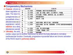
Chapter4Diseases ofcardiovascular systemVIMCompensatoryMechanisms受体类型主要分布>However,chroniceMI受体V胃壁细胞、神经节和中枢神经系统down-regulation oc受M2受体心脏、脑(间脑、脑桥)自主神经节、平滑肌state.Thereturn to体M3受体外分泌腺、平滑肌、血管内皮、脑、自主神经节juxtaglomerular近肾NNm受体神经肌肉接头受Nn受体神经节、中枢sympatheticdrive体aldosterone醛固酮α1受体血管平滑肌、瞳孔开大肌、心脏和肝脏aangiotensin I which受α2受体血管平滑肌、血小板、脂肪细胞、去甲肾上腺素能和胆碱能神体经末梢retention via a direcβ1受体心脏、肾小球旁系细胞βaldosterone secret受β2受体平滑肌、骨骼肌、肝等Ultimately,theactiv体β3受体脂肪细胞cardiac output and is anothermechanismbywhich thebodyattemptstonormalizecardiovascularfunction.Eventually,progressivemyocardialfailureand chronicactivationoftheRAASresult inpulmonaryedemaGuangdong Ocean University
7 uCompensatory Mechanisms Ø However, chronic elevation of sympathetic drive damages the myocardium and β-receptor down-regulation occurs. This returns myocardial contractility to its previously reduced state. The return to reduced cardiac output results in a decrease in sodium delivery to the juxtaglomerular近肾小球的 apparatus of the kidneys, which, along with chronic elevation of sympathetic drive, results in increased renin release, activation of renin-angiotensin- aldosterone醛固酮 system (RAAS), and conversion of angiotensinogen (liver produced to angiotensin I which is then converted to angiotensin II. This increases sodium and water retention via a direct effect on the renal tubules as well as secondarily via an increase in aldosterone secretion, to increase the blood volume. Ø Ultimately, the activation of the RAAS results in increased blood volume and increased cardiac output and is another mechanism by which the body attempts to normalize cardiovascular function. Eventually, progressive myocardial failure and chronic activation of the RAAS result in pulmonary edema
7Chapter4Diseases ofcardiovascular systemVIMIClinicalManifestations>Left-sidedCongestiveHeartFailure:Withleft-sided CHF充血性心力衰竭,clinical signs areassociated withan increase in pulmonaryvenous and capillary hydrostatic pressure. Pulmonary edema andcongestion (cough, dyspnea) are the most common signs.Right-sided Congestive Heart Failure: Right-sided CHF results inan increase in pressure in the vessels delivering blood to the rightventricle--the systemic veins and systemic capillaries. This can resultin ascites腹水, pleural effusion, and/or peripheral edema.Biventricular Failure: This can arise in patients with myocardialfailure resulting from dilated cardiomyopathy or toxin exposure.Clinical signs attributable to both forms of CHF can be noted, althoughcommonly signs of one will predominate.Guangdong Ocean University
8 nClinical Manifestations Ø Left-sided Congestive Heart Failure: With left-sided CHF充血性心力 衰竭, clinical signs are associated with an increase in pulmonary venous and capillary hydrostatic pressure. Pulmonary edema and congestion (cough, dyspnea) are the most common signs. Ø Right-sided Congestive Heart Failure: Right-sided CHF results in an increase in pressure in the vessels delivering blood to the right ventricle—the systemic veins and systemic capillaries. This can result in ascites腹水, pleural effusion, and/or peripheral edema. Ø Biventricular Failure: This can arise in patients with myocardial failure resulting from dilated cardiomyopathy or toxin exposure. Clinical signs attributable to both forms of CHF can be noted, although commonly signs of one will predominate
★Chapter4Diseases ofcardiovascularsystemVIMTherapyforheartfailure> To reduce signs of congestion (pulmonary edema, ascites,pleural effusion).Diuretics:Furosemide速尿.Chlorthiazide氯噻嗪aresometimesaddedtothetherapeuticregimenof CHFPositive Inotropes干预:The digitalisglycosides洋地黄糖苷(digoxin地高辛)arethemostcommonlyusedpositiveinotropicagents and cause a modest increase in cardiac contractility.Angiotensin-convertingEnzyme(ACE血管紧张素转化酶)Inhibitors:They have become an important tool in thetreatment of mild to severe CHF in dogs. Enalapril丙脯氨酸 shouldbeinitiatedindogsatadosageof0.5mg/kg,PO,sid-一天一次.Tobe continuedGuangdong Ocean University
9 uTherapy for heart failure Ø To reduce signs of congestion (pulmonary edema, ascites, pleural effusion). • Diuretics: Furosemide速尿. Chlorthiazide氯噻嗪 are sometimes added to the therapeutic regimen of CHF. • Positive Inotropes干预: The digitalis glycosides洋地黄糖苷 (digoxin地高辛) are the most commonly used positive inotropic agents and cause a modest increase in cardiac contractility. • Angiotensin-converting Enzyme (ACE血管紧张素转化酶) Inhibitors: They have become an important tool in the treatment of mild to severe CHF in dogs. Enalapril丙脯氨酸 should be initiated in dogs at a dosage of 0.5 mg/kg, PO, sid一天一次. To be continued
Chapter4Diseases of cardiovascular systemVIMVasodilators: Hydralazine耕苯哒嗪 is a potent arteriolar vasodilator thatdirectlydilatesarterioles.β-AdrenergicBlockers:Experimental dogs with CHF administered a β-adrenergic肾上腺素的 blocking drug (eg, propranolol心得安),Calcium Channel Blockers: Calcium channel blockers such as diltiazem盐酸地尔硫卓 are used in the treatment of heart failure secondary to hypertrophiccardiomyopathySupplementation: Dogs are treated with taurine牛磺酸 500-1,000 mg, PO,bid.The dosage in cats is 250 mg, PO, bid.Low-sodium Diets.Oxygen Therapy: The percent inspired oxygen is generally increased to 4050-100%Abdominocentesis腹腔穿刺术: In dogs and cats with severe right-sided CH, ascites can besevere enough to result in dyspnea. Abdominocentesis is a safe and effective means oftreating this fluid accumulation and can be performed on a regular basis(every2-4wk if needed)as long as thepatient is cooperative.10Guangdong Ocean University
10 • Vasodilators: Hydralazine肼苯哒嗪 is a potent arteriolar vasodilator that directly dilates arterioles. • β-Adrenergic Blockers: Experimental dogs with CHF administered a β- adrenergic肾上腺素的 blocking drug (eg, propranolol心得安). • Calcium Channel Blockers: Calcium channel blockers such as diltiazem盐酸地 尔硫卓 are used in the treatment of heart failure secondary to hypertrophic cardiomyopathy. • Supplementation: Dogs are treated with taurine牛磺酸 500-1,000 mg, PO, bid. The dosage in cats is 250 mg, PO, bid. • Low-sodium Diets. • Oxygen Therapy: The percent inspired oxygen is generally increased to 40- 50-100%. • Abdominocentesis腹腔穿刺术: In dogs and cats with severe right-sided CHF, ascites can be severe enough to result in dyspnea. Abdominocentesis is a safe and effective means of treating this fluid accumulation and can be performed on a regular basis (every 2-4 wk if needed) as long as the patient is cooperative
按次数下载不扣除下载券;
注册用户24小时内重复下载只扣除一次;
顺序:VIP每日次数-->可用次数-->下载券;
- 广东海洋大学:《兽医内科学》课程教学课件(讲稿)Chapter 3 Diseases of Respiratory System.pdf
- 广东海洋大学:《兽医内科学》课程教学课件(讲稿)Chapter 2 Diseases of Digestive System.pdf
- 广东海洋大学:《兽医内科学》课程教学课件(讲稿)Chapter 1 Introduction to VIM.pdf
- 广东海洋大学:《兽医内科学》课程教学资源(教案讲义,共六章,任课教师:陈进军、康丹菊).doc
- 广东海洋大学:《兽医内科学》实验课程教学大纲 Veterinary Internal Medicine.doc
- 广东海洋大学:《兽医内科学》课程实验指导(讲义)实验四 血液丙酮酸的测定.doc
- 广东海洋大学:《兽医内科学》课程实验指导(讲义)实验三 血清谷丙、谷草转氨酶活力测定(赖氏法).doc
- 广东海洋大学:《兽医内科学》课程实验指导(讲义)实验二 氢氰酸的定性与定量检验.doc
- 广东海洋大学:《兽医内科学》课程实验指导(讲义)实验一 血清总钙含量的测定.doc
- 塔里木大学:《兽医临床诊断学》课程实验课教案.docx
- 塔里木大学:《兽医临床诊断学》课程实验教学大纲.pdf
- 塔里木大学:《兽医临床诊断学》课程理论课教案.docx
- 塔里木大学:《兽医临床诊断学》课程教学大纲 Veterinary Clinical Diagnostics.pdf
- 《兽医外科学》课程教学课件(PPT讲稿)第十五章 小动物牙病.ppt
- 《兽医外科学》课程教学课件(PPT讲稿)第十四章 四肢疾病.ppt
- 《兽医外科学》课程教学课件(PPT讲稿)第十三章 跛行诊断.ppt
- 《兽医外科学》课程教学课件(PPT讲稿)第十二章 泌尿生殖系统疾病.ppt
- 《兽医外科学》课程教学课件(PPT讲稿)第十一章 直肠及肛门疾病.ppt
- 《兽医外科学》课程教学课件(PPT讲稿)第七章 头部疾病.ppt
- 《兽医外科学》课程教学课件(PPT讲稿)第八章 枕、颈部疾病.ppt
- 广东海洋大学:《兽医内科学》课程教学课件(讲稿)Chapter 5 Diseases of Urinary system.pdf
- 广东海洋大学:《兽医内科学》课程教学课件(讲稿)Chapter 6 Diseases of the nervous system.pdf
- 西昌学院:《动物遗传学》实验课程教学大纲.docx
- 西昌学院:《家畜解剖学》实验课程教学指导(共十八个实验).doc
- 龙岩学院:《预防兽医学》实验课程教学大纲.docx
- 龙岩学院:《基础兽医学》实验课程教学大纲.docx
- 龙岩学院:《临床兽医学》实验课程教学大纲.docx
- 南京农业大学:《兽医内科学》实验课程教学大纲(强化).doc
- 南京农业大学:《兽医内科学》实验课程教学大纲(动医).doc
- 南京农业大学:《兽医内科学》实验课程教学大纲.doc
- 南京农业大学:《兽医临床诊断学》实验课程教学大纲(动医、实验).doc
- 南京农业大学:《动物解剖学》课程II教学大纲(动医).pdf
- 南京农业大学:《动物解剖学》课程II教学大纲(动物实验).pdf
- 南京农业大学:《宠物解剖学》课程教学大纲.pdf
- 南京农业大学:《中兽医学》实验课程教学大纲.pdf
- 南京农业大学:《兽医药理学》实验课程教学大纲.pdf
- 南京农业大学:《兽医药代动力学》实验课程教学大纲.pdf
- 南京农业大学:《药物化学》实验课程教学大纲.pdf
- 南京农业大学:《兽医毒理学》实验课程教学大纲.pdf
- 南京农业大学:《药物分析》实验课程教学大纲.pdf
