同济大学:《生理学》课程电子教案(PPT课件)03 Basics of the ElectroCardioGram(ECG)
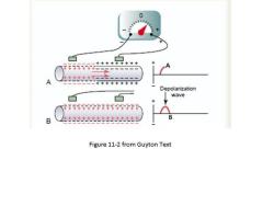
0 +++++++++ Depolarization 白 wave 行行行用 B Figure 11-2 from Guyton Text
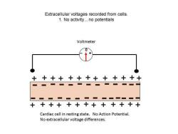
Extracellular voltages recorded from cells. 1.No activity...no potentials Voltmeter ++++++++++++ 十 ■ ■一■■■■■■ +++++++++++++ Cardiac cell in resting state.No Action Potential. No extracellular voltage differences
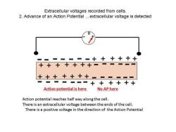
Extracellular voltages recorded from cells. 2.Advance of an Action Potential...extracellular voltage is detected ▣===+十十+十十 十++++十+■一==▣▣ 十十十十十十十====== ■-■■■一■十++十+十 Action potential is here No AP here Action potential reaches half way along the cell. There is an extracellular voltage between the ends of the cell. There is a positive voltage in the direction of the Action Potential
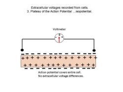
Extracellular voltages recorded from cells. 3.Plateau of the Action Potential...isopotential. Voltmeter 0 ■=■=一■■==■■一 ++十++++++++++ +++++++十+++++ Action potential covers entire cell. No extracellular voltage differences
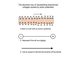
The standard way of representing extracellular voltages caused by action potentials: ▣=一=▣■▣++十+十十 十++十十+十=一一一一▣ 十++十十十+▣-■✉= ■■■■=■+十++十十 1.Here is a cell with an action potential. X 2.Represent the cell as a dipole 3.Use an arrow to denote the polarity of the dipole
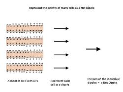
Represent the activity of many cells as a Net Dipole +++++t+特 ■一■■=■+十十+++ ==■=■■++十+++ +++++++一一一一一▣ +++++++ ±共++±+中++++ ■=+十十++十 +++++干+合 ■■=■■■+十十+十十 The sum of the individual A sheet of cells with APs Represent each dipoles a Net Dipole cell as a dipole
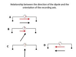
Relationship between the direction of the dipole and the orientation of the recording axis
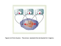
0 Figure 11-4 from Guyton.The arrows represent the net dipoles for 2 regions
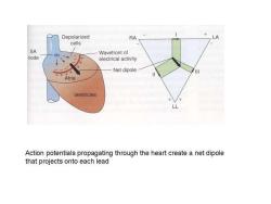
Depolarized RA LA cells SA Wavefront of node electrical activity Net dipole Atria Ventricles L Action potentials propagating through the heart create a net dipole that projects onto each lead
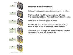
SA node 0 Sequence of activation of heart. Cells activated by action potentials are depicted in yellow. 2 The first cells to reach threshold are in the SA node APs are conducted to the AV node through atrial myocytes. anter Conduction is slow through the AV node. From the AV node APs are conducted through Purkinje fibers(Bundle of His)to the bottom of the ventricles. myocard,froe The bundle splits into right and left branches and activates myocytes in the right and left ventricles
按次数下载不扣除下载券;
注册用户24小时内重复下载只扣除一次;
顺序:VIP每日次数-->可用次数-->下载券;
- 同济大学:《生理学》课程电子教案(PPT课件)02 Cardiac Action Potential.ppt
- 同济大学:《生理学》课程电子教案(PPT课件)01 Overview of Cardiovascular System(负责人:汪海宏).ppt
- 同济大学:《生理学》课程教学资源(试卷习题)试卷01.pdf
- 新乡医学院:《毒理学》课程教学资源(课件讲稿,打印版)实验六 安全性评价.pdf
- 新乡医学院:《毒理学》课程教学资源(教案讲义,打印版)实验六 安全性评价.pdf
- 新乡医学院:《毒理学》课程教学资源(课件讲稿,打印版)实验五 血清谷丙转氨酶活性的测定(King法).pdf
- 新乡医学院:《毒理学》课程教学资源(教案讲义,打印版)实验五 实验动物染毒.pdf
- 新乡医学院:《毒理学》课程教学资源(课件讲稿,打印版)实验四 急性经口毒性实验.pdf
- 新乡医学院:《毒理学》课程教学资源(教案讲义,打印版)实验四 经口急性毒性实验.pdf
- 新乡医学院:《毒理学》课程教学资源(课件讲稿,打印版)实验三 生物材料的采集与制备.pdf
- 新乡医学院:《毒理学》课程教学资源(课件讲稿,打印版)实验二 实验动物染毒(染毒方法介绍).pdf
- 新乡医学院:《毒理学》课程教学资源(教案讲义,打印版)实验三 生物材料的采集.pdf
- 新乡医学院:《毒理学》课程教学资源(课件讲稿,打印版)实验一 动物基本知识及基本操作、实验方法.pdf
- 新乡医学院:《毒理学》课程教学资源(教案讲义,打印版)实验二 实验动物染毒.pdf
- 新乡医学院:《毒理学》课程教学资源(教案讲义,打印版)实验一 动物基本知识及基本操作、实验方法.pdf
- 《病原生物学与免疫学》课程教学资源(教材讲义)病原生物学与免疫学参考教材PDF电子版(高职,共二十六章).pdf
- 安庆医药高等专科学校:《病原生物与免疫学》课程教学资源(PPT课件)绪论——免疫与医学免疫学概述.ppt
- 安庆医药高等专科学校:《病原生物与免疫学》课程教学资源(PPT课件)高中病免知识.ppt
- 安庆医药高等专科学校:《病原生物与免疫学》课程教学资源(PPT课件)病原生物学概述.ppt
- 安庆医药高等专科学校:《病原生物与免疫学》课程教学资源(PPT课件)第二十五章 医学原虫(叶足虫-溶组织内阿米巴).ppt
- 同济大学:《生理学》课程教学资源(试卷习题)生理学多选题(一).pdf
- 同济大学:《生理学》课程教学资源(试卷习题)生理学多选题(三).pdf
- 同济大学:《生理学》课程教学资源(试卷习题)生理学多选题(二).pdf
- 同济大学:《生理学》课程教学资源(试卷习题)试卷02.pdf
- 同济大学:《生理学》课程电子教案(PPT课件)04 Excitation-Contraction Coupling in Cardiac Cells.ppt
- 同济大学:《生理学》课程电子教案(PPT课件)05 Cardiac Muscle Mechanics.ppt
- 《抗体及其应用》课程教学资源(参考资料,电子版)01-Antibody.pdf
- 《抗体及其应用》课程教学资源(参考资料,电子版)02-Monoclonal antibody.pdf
- 《抗体及其应用》课程教学资源(参考资料,电子版)03-Monoclonal antibody therapy.pdf
- 《抗体及其应用》课程教学资源(参考资料,电子版)04-Cancer immunotherapy.pdf
- 《抗体及其应用》课程教学资源(参考资料,电子版)05-Immunoassay.pdf
- 《抗体及其应用》课程教学资源(参考资料,电子版)06-Antibody microarray.pdf
- 广东医科大学:《抗体及其应用》课程教学资源(课件讲稿,打印版)01-抗体的结构与功能.pdf
- 广东医科大学:《抗体及其应用》课程教学资源(课件讲稿,打印版)02-多克隆抗体.pdf
- 广东医科大学:《抗体及其应用》课程教学资源(课件讲稿,打印版)03-单克隆抗体的制备.pdf
- 广东医科大学:《抗体及其应用》课程教学资源(课件讲稿,打印版)04-基因工程抗体之嵌合抗体.pdf
- 广东医科大学:《抗体及其应用》课程教学资源(课件讲稿,打印版)05-抗体的应用.pdf
- 广东医科大学:《人体解剖学》课程实验指导(打印版)局部解剖学实验指导.pdf
- 广东医科大学:《人体解剖学》课程实验指导(打印版)实验指导及强化训练习题集及答案(适用于医学本科临床、法医等专业).pdf
- 广东医科大学:《生理学》课程教学资源(大纲教案)生理学教学大纲 Physiology.pdf
