重庆医科大学:《病理学》课程英文试卷(答案)

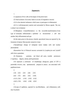
Answers 1. (1)apoptosis of liver cells at the margin of the portal tracts (2)Total occlusion of an artery leads to an area of coagulative necrosis (3)It is the chemical substances which cause or participate in inflammation (4)It is well-demarcated casetion and surrounded by fibrous capsule.The size is about 3 cm in diameter (5)Metaplasia (transdiffentiation)is the reversible transformation of one type of terminally differentiated epithelial or messenchmal )cell into another fully differentiated cell type (6)the name given to the process whereby specialized tissue are repaired by the formation of mature fibrovascular connective tissue (7)morphologic change of malignant tumor display cells and nuclear pleomophism. (8)constitute foci of fibrinoid necrosis surrouded by lymphocytes and Aschoff cells form a granulama (9)Hematuria azotemia,red blood cell casts,variable proteinuria (<Igm/day),oliguria,edema,and hypertension (10)represent a continuum of morphologic change:any grade of CIN is potentially invasive.may spontaneously progress to cancer:are associated with papillomaviruses 2 (1)F;(2)T(3)T(4)F;(5)T (6)T(7)T(8)T(9)T(10F (11)T(12)F;(13)T(14)T(15)T (16)T(17)F,(18)T(19)F;(20)T 3. (1)Loops of capillaries.suported by myofibroblastes.and inflammatory cells may be present
Answers 1. (1) apoptosis of liver cells at the margin of the portal tracts (2) Total occlusion of an artery leads to an area of coagulative necrosis (3) It is the chemical substances which cause or participate in inflammation (4) It is well-demarcated casetion and surrounded by fibrous capsule. The size is about 3 cm in diameter (5) Metaplasia ( transdiffentiation ) is the reversible transformation of one type of terminally differentiated ( epithelial or messenchmal ) cell into another fully differentiated cell type. (6) the name given to the process whereby specialized tissue are repaired by the formation of mature fibrovascular connective tissue (7)morphologic change of malignant tumor display cells and nuclear pleomophism. (8)constitute foci of fibrinoid necrosis surrouded by lymphocytes and Aschoff cells form a granulama (9) Hematuria. azotemia, red blood cell casts, variable proteinuria (<1gm/day), oliguria, edema, and hypertension (10) represent a continuum of morphologic change;any grade of CIN is potentially invasive, may spontaneously progress to cancer ; are associated with papillomaviruses. 2. (1)F; (2)T; (3)T; (4)F; (5)T; (6)T; (7)T; (8)T; (9)T; (10)F; (11)T; (12)F; (13)T; (14)T; (15)T; (16)T; (17)F; (18)T; (19)F; (20)T. 3. (1)Loops of capillaries, suported by myofibroblastes, and inflammatory cells may be present

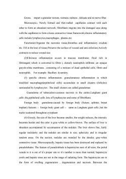
Gross:impart a granular texture,wetness,redness,delicate and no nerve fiber. Microscopic:Newly formed and thin-walled capillaries connect with each other to form an abundant network,fibroblasts migrate into the damaged area along with the capillaries to form a loose connective tissue framework.chronic inflammatory cells include lymphocytes,macrophages,plasma etc. Functiones:Organise the necrostic tissue,thrombus and inflammatory exudate etc.Fill in the lose of tissue Preserve the surface of wound and anti-infectionActively contracts to reduce wound size (2)Fibrinous inflammation occurs in mucous membrane;fluid rich in fibrinogen(which is converted to fibrin )densely neutrophils infiltrate an opaque greysh-white membrane,consisting of a mixture of dead epithelial cells,fibrin and neutrophils.For example:Bacillary dysentery. (3)specific chronic inflammation:granulomatous inflammation in which modified macrophages(epithelioid cells)accumulate in small clusters (follicles) surrounded by lymphocytes.The small clusters are called granulomas Guanuloma of tuberculosis:caseous necrosis in the centre;Langhans giant cells :the epithelioid cellslots of lymphocytes and some of fibroblasts. Foreign body granuloma:caused by foreign body (Suture,splinter,breast implant)features foreign body giant cell-same as Langhans giant cells,but the nuclei scattered throughout cytoplasm (4)Grossly,the size of the liver become smaller,the weight reduces,the intensity becomes harder and the color is gray-white or yellow-brown.The surface of liver is shrunken accompanied by accentuation of the nodules.The liver shows fine,fairly regular nodularity and the nodules are similar in size,sphericity and in irregular random areas.On the section,nodules are rounded by the slender,gray-white connective tissue.Microscopically,hepatic tissue has been destroyed and replaced by pseudolobules.The feature of pseudolobule is hepatocytes nest of all sizes,the portal venule in it is out of it's proper site or it's number is more than normal,hepatocyte cords and hepatic sinus are not in the range of radiating form.The hepatocytes are in the form of swelling ,regeneration,degeneration and necrosis.Between the
Gross: impart a granular texture, wetness, redness , delicate and no nerve fiber. Microscopic:Newly formed and thin-walled capillaries connect with each other to form an abundant network; fibroblasts migrate into the damaged area along with the capillaries to form a loose connective tissue framework;chronic inflammatory cells include lymphocytes,macrophages, plasma etc. Functiones:Organise the necrostic tissue,thrombus and inflammatory exudate etc. Fill in the lose of tissue;Preserve the surface of wound and anti-infection;Actively contracts to reduce wound size (2)Fibrinous inflammation occurs in mucous membrane; fluid rich in fibrinogen( which is converted to fibrin ), densely neutrophils infiltrate ;an opaque greysh-white membrane , consisting of a mixture of dead epithelial cells, fibrin and neutrophils. For example: Bacillary dysentery. (3) specific chronic inflammation: granulomatous inflammation in which modified macrophages(epithelioid cells) accumulate in small clusters (follicles) surrounded by lymphocytes . The small clusters are called granulomas Guanuloma of tuberculosis:caseous necrosis in the centre;Langhans giant cells ;the epithelioid cells ;lots of lymphocytes and some of fibroblasts. Foreign body granuloma:caused by foreign body (Suture, splinter, breast implant) features :foreign body giant cell - same as Langhans giant cells ,but the nuclei scattered throughout cytoplasm (4) Grossly, the size of the liver become smaller, the weight reduces, the intensity becomes harder and the color is gray-white or yellow-brown. The surface of liver is shrunken accompanied by accentuation of the nodules. The liver shows fine, fairly regular nodularity and the nodules are similar in size, sphericity and in irregular random areas. On the section, nodules are rounded by the slender, gray-white connective tissue. Microscopically, hepatic tissue has been destroyed and replaced by pseudolobules. The feature of pseudolobule is hepatocytes nest of all sizes, the portal venule in it is out of it’s proper site or it’s number is more than normal, hepatocyte cords and hepatic sinus are not in the range of radiating form. The hepatocytes are in the form of swelling ,regeneration , degeneration and necrosis. Between the

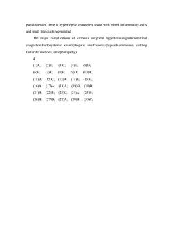
pseudolobules,there is hypertrophic connective tissue with mixed inflammatory cells and small bile ducts regenerated. The major complications of cirrhosis are:portal hypertension(gastrointestinal congestion,Portosystemic Shunts).hepatic insufficiency(hypoalbuminaemia,clotting factor deficiencies,encephalopathy) 4. (I)A;(2)E;(3)C;(4)E;(5)D (6)E(7)E;(8)E:(9D:(10)A: (11)B,(12)C,(13)A:(14)E,(15)E: (16)A:(17)A;(18)A;(19)B:(20)B: (21)B(22)B,(23)C,(24)A;(25)B (26)B:(27)D:(28)A,(29)B(30)C
pseudolobules, there is hypertrophic connective tissue with mixed inflammatory cells and small bile ducts regenerated . The major complications of cirrhosis are:portal hypertension(gastrointestinal congestion,Portosystemic Shunts);hepatic insufficiency(hypoalbuminaemia, clotting factor deficiencies, encephalopathy) 4. (1)A; (2)E; (3)C; (4)E; (5)D; (6)E; (7)E; (8)E; (9)D; (10)A; (11)B; (12)C; (13)A: (14)E; (15)E; (16)A; (17)A; (18)A; (19)B; (20)B; (21)B; (22)B; (23)C; (24)A; (25)B; (26)B; (27)D; (28)A; (29)B; (30)C;
按次数下载不扣除下载券;
注册用户24小时内重复下载只扣除一次;
顺序:VIP每日次数-->可用次数-->下载券;
- 重庆医科大学:《病理学》课程英文试卷(试题).doc
- 重庆医科大学:《病理学》课程作业习题(英文,无答案).doc
- 重庆医科大学:《病理学》课程教学实习指导(英文).doc
- 重庆医科大学:《病理学》课程课程教学大纲 A Teaching Outline for Pathology Course.doc
- 重庆医科大学:《病理学》课程理论教学大纲(供七年制和五年制使用).doc
- 重庆医科大学:《病理学》课程教学资源(PPT课件)传染病与寄生虫病(infectious desease and parasitosis).ppt
- 重庆医科大学:《病理学》课程教学资源(PPT课件)神经系统疾病(Diseases of the CNS).ppt
- 重庆医科大学:《病理学》课程教学资源(PPT课件)内分泌系统疾病.ppt
- 重庆医科大学:《病理学》课程教学资源(PPT课件)女性生殖系统疾病(disease of female reproductive system and breast).ppt
- 重庆医科大学:《病理学》课程教学资源(PPT课件)泌尿系统疾病 Diseases of urinary system.ppt
- 重庆医科大学:《病理学》课程教学资源(PPT课件)消化系统疾病 Digestive system disease.ppt
- 重庆医科大学:《病理学》课程教学资源(PPT课件)呼吸系统疾病.ppt
- 重庆医科大学:《病理学》课程教学资源(PPT课件)心血管疾病 Diseases of Cardiovascular system.ppt
- 重庆医科大学:《病理学》课程教学资源(PPT课件)肿瘤(tumor, neoplasm).ppt
- 重庆医科大学:《病理学》课程教学资源(PPT课件)炎症 inflammation.ppt
- 重庆医科大学:《病理学》课程教学资源(PPT课件)局部血液循环障碍.ppt
- 重庆医科大学:《病理学》课程教学资源(PPT课件)绪论、细胞和组织的适应与损伤、损伤的修复.ppt
- 《病理学》课程作业习题(含参考答案).pdf
- 《病理学》课程实习指导 Guidance of Pathology.pdf
- 《病理学》课程教学大纲(五年制临床、儿科、麻醉、基础、预防、口腔医学专业使用;七年制临床、儿科、检验、物理专业使用).docx
- 重庆医科大学:《病理学》课程教学资源(授课教案)infectious disease.doc
- 重庆医科大学:《病理学》课程教学资源(授课教案)the Diseases of female genital system.doc
- 重庆医科大学:《病理学》课程教学资源(授课教案)disease of nevous system_1.doc
- 重庆医科大学:《病理学》课程教学资源(授课教案)disease of nevous system_2.doc
- 重庆医科大学:《病理学》课程教学资源(授课教案)diseases of cardiovascular system.doc
- 重庆医科大学:《病理学》课程教学资源(授课教案)diseases of respiratory system.doc
- 重庆医科大学:《病理学》课程教学资源(授课教案)disease of alimentary system.doc
- 重庆医科大学:《病理学》课程教学资源(授课教案)disease of urinary system.doc
- 重庆医科大学:《病理学》课程教学资源(授课教案)cell adaptation and injury, repair.doc
- 重庆医科大学:《病理学》课程教学资源(授课教案)tumor.doc
- 重庆医科大学:《病理学》课程教学资源(授课教案)Introduction and hemodynamic rearangment.doc
- 重庆医科大学:《病理学》课程教学资源(授课教案)inflammation.doc
- 重庆医科大学:《病理学》课程教学资源(授课教案)内分泌系统疾病.doc
- 重庆医科大学:《病理学》课程教学资源(授课教案)传染和寄生虫病.doc
- 重庆医科大学:《病理学》课程教学资源(授课教案)女性生殖系统疾病.doc
- 重庆医科大学:《病理学》课程教学资源(授课教案)神经系统疾病.doc
- 重庆医科大学:《病理学》课程教学资源(授课教案)心血管系统疾病.doc
- 重庆医科大学:《病理学》课程教学资源(授课教案)呼吸系统疾病.doc
- 重庆医科大学:《病理学》课程教学资源(授课教案)消化系统疾病.doc
- 重庆医科大学:《病理学》课程教学资源(授课教案)泌尿系统疾病.doc
