《儿科学》课程作业习题(典型病例)03 congenital heart disease-VSD

Medical number: 668197Diagnosis: ventricular septal defect (VSD)History summary:Male, 2 years and 6 monthChief complaints: post-c-exercisetachypneafor2yearswhile feeding. Heachypnegoespiratoryearsinfection since then. 114 month ago, he was taken to hospital when having a cold Pediatricianheard heart murmur during physical examination. In our hospital, he was diagnosed as ventricularseptal defect by echocardiography. The parents didn't complain the signs of cyanosis. squattingncope.diaphoSisorpoorweightgalThe child was full-term.Themother was healthy during pregnancyPasthistoryImmunizations: All.Feeding.GoodFamily History:THarentsarehealthy.The family has no history of congenital healiseasion: T 36.6℃, P:113bpm, R: 34bpm,Phvsidmisweight:14kg,BP94/65mmHg.Hewases or edema. No moist and dry rales ins and quiet.No cyanosisashboth lungs. Heart: no visible lifts. Systolic thrill was palpable at the lower left sternal borderbetween the third and fourth intercostals spaces. A grade II/VI, harsh pansystolic murmur washeard at this area The second sound at the pulmonary area was normal.Abdomen: Soft.no painmass,thill orfluid wave.Liver is palpableIcmbelow thecostal marginNo clubbing offingers angInvestigation test:(I) Echocardiogram: LA and LV enlargement. VSD, PFO, Mild MR, TR, PR.(2) ECG: normal.(3) Chest radiograph: Increase pulmonaryvasculature.TreatmenTrans-catheter VSD occlusion was performed
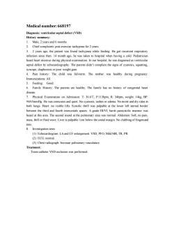
Medical number: 668197 Diagnosis: ventricular septal defect (VSD) History summary: 1. Male, 2 years and 6 months. 2. Chief complaints: post-exercise tachypnea for 2 years. 3. 2 years ago, the patient was found tachypnea while feeding. He got recurrent respiratory infection since then. 14 month ago, he was taken to hospital when having a cold. Pediatrician heard heart murmur during physical examination. In our hospital, he was diagnosed as ventricular septal defect by echocardiography. The parents didn’t complain the signs of cyanosis, squatting, syncope, diaphoresis or poor weight gain. 4. Past history: The child was full-term. The mother was healthy during pregnancy. Immunizations: All. 5. Feeding: Good. 6. Family History: The parents are healthy. The family has no history of congenital heart disease. 7. Physical Examination on Admission: T: 36.6℃, P:113bpm, R: 34bpm, weight: 14kg, BP: 94/65mmHg. He was conscious and quiet. No cyanosis, rashes or edema. No moist and dry rales in both lungs. Heart: no visible lifts. Systolic thrill was palpable at the lower left sternal border between the third and fourth intercostals spaces. A grade III/VI, harsh pansystolic murmur was heard at this area. The second sound at the pulmonary area was normal. Abdomen: Soft, no pain, mass, thill or fluid wave. Liver is palpable 1cm below the costal margin. No clubbing of fingers and toes. 8. Investigation tests: (1) Echocardiogram: LA and LV enlargement. VSD, PFO, Mild MR, TR, PR. (2) ECG: normal. (3) Chest radiograph: Increase pulmonary vasculature. Treatment: Trans-catheter VSD occlusion was performed
按次数下载不扣除下载券;
注册用户24小时内重复下载只扣除一次;
顺序:VIP每日次数-->可用次数-->下载券;
- 《儿科学》课程作业习题(典型病例)08 diarrhea.doc
- 《儿科学》课程作业习题(典型病例)06 anute nepheritis-1.doc
- 《儿科学》课程作业习题(典型病例)06 nephrotic syndrome-2.doc
- 《儿科学》课程作业习题(复习题)01 questions of children healthcare.doc
- 《儿科学》课程作业习题(复习题)02 questions of neonatal diseases.doc
- 《儿科学》课程作业习题(复习题)05 questions of blood disorders.doc
- 《儿科学》课程作业习题(复习题)04 questions of circulatory system.doc
- 《儿科学》课程作业习题(复习题)03 questions of respiratory diseases.doc
- 《儿科学》课程作业习题(复习题)06 questions of nervous system.doc
- 《儿科学》课程作业习题(复习题)10 questions of infectious diseases.doc
- 《儿科学》课程作业习题(复习题)09 questions of endocrine disorders.doc
- 《儿科学》课程作业习题(复习题)07 questions of urinological system.doc
- 《儿科学》课程作业习题(复习题)08 questions of immune system.doc
- 《儿科学》课程教学资源(授课教案)12 Acute Glomerulonephritis,Nephrotic Syndrome.doc
- 《儿科学》课程教学资源(授课教案)11 Acute Convulsion in Children.doc
- 《儿科学》课程教学资源(授课教案)13 Immunodeficiency.doc
- 《儿科学》课程教学资源(授课教案)10 Nutritional Iron Deficiency Anemia.doc
- 《儿科学》课程教学资源(授课教案)15 Growth Hormone Deficiency.doc
- 《儿科学》课程教学资源(授课教案)14 Congenital Hypothyroidism.doc
- 《儿科学》课程教学资源(授课教案)17 Varicella.doc
- 《儿科学》课程作业习题(典型病例)04 iron deficiency anemia.doc
- 《儿科学》课程作业习题(典型病例)05 purulent meningitis.doc
- 《儿科学》课程作业习题(典型病例)07 congenital hypothyroidism.doc
- 《儿科学》课程作业习题(典型病例)03 congenital heart disease-TOF.doc
- 《儿科学》课程作业习题(典型病例)01 ABO incompatibility of neonates.doc
- 《儿科学》课程作业习题(典型病例)02 pneumonia.doc
- 《儿科学》课程作业习题(试卷和答案)双语试卷C卷(试题).doc
- 《儿科学》课程作业习题(试卷和答案)双语试卷B卷(试题).doc
- 《儿科学》课程作业习题(试卷和答案)双语试卷B卷(答案).doc
- 《儿科学》课程作业习题(试卷和答案)双语试卷C卷(答案).doc
- 《儿科学》课程作业习题(试卷和答案)双语试卷A卷(试题).doc
- 《儿科学》课程作业习题(试卷和答案)双语试卷A卷(答案).doc
- 《儿科学》课程教学课件(PPT讲稿)07 新生儿缺氧缺血性脑病 Hypoxic-ischemic Encephalopathy(HIE).pptx
- 《儿科学》课程教学课件(PPT讲稿)06 新生儿败血症 Neonatal Septicemia.pptx
- 《儿科学》课程教学课件(PPT讲稿)21 Chronic Gastritis in Children.pptx
- 《儿科学》课程教学课件(PPT讲稿)20 Toxic Bacillary Dysentery.pptx
- 《儿科学》课程教学课件(PPT讲稿)27 Inflammation Causes Cholesterol Redistribution by Diverting Cholesterol from Circulation to Tissue Tompartments.pptx
- 《儿科学》课程教学课件(PPT讲稿)26 Rotavirus Infection in Children.pptx
- 《儿科学》课程教学课件(PPT讲稿)25 Scarlet Fever.pptx
- 《儿科学》课程教学课件(PPT讲稿)24 Mumps(Epidemic parotitis).pptx
