《儿科学》课程作业习题(典型病例)05 purulent meningitis

Medical number:658527Main diagnosis:1.Acute Bacterial Meningitis2. Subdural effusionsHydrocephalusSepticemiaHistory:Boy,5months.2.Main complaints:feverfor 5days and lethargy for 4 days.Hehad feverfor5days beforehewas admitted to in-patient department of our hospital.Thewas high as39.2CFourdays ago,hebecamepoorfeeding and irritation Aftertemperatuthat, he had tonic-clonic seizures. He had no bulging fontanel, had no disturbance ofconsciousness. During the course, he had dirrhea, but no vomit, no rush, no cough orwheezing, no jaundice. Afer regular treatment with antibiotics in our out-patient departmentfor 5 days, thesno improvement with his symptomVaccilvaccineafter birthHehadBCGPast Histohistoryof contact with TB.,no historyof otitismediaPhysical Exams(1) General Exam: T39.2°C, P172/min, R 43/min Poor appearance, awake and iritable. Theface was pale without cyanosis. Anterior fontanel was nearly closed with normal tension.with good air entry throughout. No wheezes, crackles, or stridor.Lungsclear toauscultatCardAbdonsoft abdomen with no palpable masswas 2.5 cm and 2.0cm under the right costal margin along the mid-clavicular line and xiphoidresptively,Spleen was not palpated(2) Neurologic Exam: awake, and irritable Cranial nerves: nomal. Motor: normal tone, bulk, andmities.Reflexes:normaldeeptendonreflexesbilaterallyinupperandrengthinallextremmeningeal irritationNeck stiffness, Kernig's sign andoweSignsofitiesBrudzinsski's sign were negative.Babinki's sign was negative7. Lab Exams(I) Blood cell count: WBC 14.77*10/L, N 67.9%, CRP 27mg/dL.(2)Sputumculture:Streptococcuspneumonia(3)Blood cccuspneumor(4) CSF (2011-1-11): cloudy appearance, WBC 3074*10%/L, neutrophilic(79%), Glucose level was 2.21mmol/L (concomitant blood glucose level 4.7mmol/l),Protein levelwas 2.37g/L.Nobacteriawere isolated fromthe CSF(5) CSF (2011-1-21): clear appearance, WBC 24*10%/L, neutrophilic 11/24, Glucose levelwas2.97mmol/L(coomitant blood glucose level 5.7mmol/l),Protein level was 1.46g/LNoisolated from the CS(6) CSF (2011-1-27): clear appearance, WBC 4*10%/L, Glucose level was 3.19mmol/L(concomitant blood glucose level 5.22mmol/l),Proteinlevel was 0.38g/L.No bacteriawere isolatedfrom theCSF(7)Suuralfuid:WBC301/Lwih 42%NC.Proteinlevel was437g/Glucslevelwas0.05mmol/l.Nobacteria wereisolatedfromtheCSF
Medical number:658527 Main diagnosis: 1. Acute Bacterial Meningitis 2. Subdural effusions 3. Hydrocephalus 4. Septicemia History: 1. Boy, 5 months. 2. Main complaints: fever for 5 days and lethargy for 4 days. 3. He had fever for 5 days before he was admitted to in-patient department of our hospital. The temperature was high as 39.2℃. Four days ago, he became poor feeding and irritation. After that, he had tonic-clonic seizures. He had no bulging fontanel, had no disturbance of consciousness. During the course, he had diarrhea, but no vomit, no rush, no cough or wheezing, no jaundice. After regular treatment with antibiotics in our out-patient department for 5 days, there was no improvement with his symptoms. 4. Vaccinations: He had BCG vaccine after birth. 5. Past History: No history of contact with T.B., no history of otitis media. 6. Physical Exams: (1) General Exam: T 39.2℃, P 172/min, R 43/min. Poor appearance, awake and irritable. The face was pale without cyanosis. Anterior fontanel was nearly closed with normal tension. Lungs: clear to auscultation, with good air entry throughout. No wheezes, crackles, or stridor. Cardiac: regular rate and rhythm. Abdomen: soft abdomen with no palpable masses. Liver was 2.5 cm and 2.0 cm under the right costal margin along the mid-clavicular line and xiphoid respectively, Spleen was not palpated. (2) Neurologic Exam: awake, and irritable. Cranial nerves: normal. Motor: normal tone, bulk, and strength in all extremities. Reflexes: normal deep tendon reflexes bilaterally in upper and lower extremities. Signs of meningeal irritation: Neck stiffness, Kernig’s sign and Brudzinski’s sign were negative. Babinki’s sign was negative. 7. Lab Exams: (1) Blood cell count: WBC 14.77*109 /L, N 67.9%, CRP 27mg/dL. (2) Sputum culture: Streptococcus pneumonia. (3) Blood culture: Streptococcus pneumonia. (4) CSF (2011-1-11): cloudy appearance, WBC 3074*106 /L, neutrophilic predominance (79%), Glucose level was 2.21mmol/L (concomitant blood glucose level 4.7mmol/l), Protein level was 2.37g/L. No bacteria were isolated from the CSF. (5) CSF (2011-1-21): clear appearance, WBC 24*106 /L, neutrophilic 11/24, Glucose level was 2.97mmol/L (concomitant blood glucose level 5.7mmol/l), Protein level was 1.46g/L. No bacteria were isolated from the CSF. (6) CSF (2011-1-27): clear appearance, WBC 4*106 /L, Glucose level was 3.19mmol/L (concomitant blood glucose level 5.22mmol/l), Protein level was 0.38g/L. No bacteria were isolated from the CSF. (7) Subdural fluid: WBC 1310*106 /L, with 42% PNC. Protein level was 4.37g/L, Glucose level was 0.05mmol/l. No bacteria were isolated from the CSF

(8) Head CT bilateral temporal and parietal subdural effusions(9) Head MRI: hydrocephalusTreatment:The boy wasgivenVancomycin270mg/d and Ceftizedime1.Og/dIVto control infection,andintravenous Mannitol todecrease ICP.On the 3day of hospitalization, the temture wasnoso highill lowfever or hypothermia sometimes.And the boy had frequent seizureTherewhich developed from lethargy to coma Head CT revealed bilateral temporalparietasubdural effusions (Figure 1).Herwent a subdural puncture.During the treattment, the boyhadmoreandmorefrequseizures,andseveredyspnea,thenhewassenttoPICUforfurthtreatment,andwasgivenVancomycin360mg/d andPanipenem500mg/dIV.On the1ith day,the'SEngedbetterasabove(2011-1-2vulsionbutseti鸣and horizontal nystagmus were noted. Head MRIshowed hydrocephalus (Figure 2) Then he underwent Ommaya reservoir implantation in the leftcerebral ventricle, and CFS was drainaged 200-300ml per day after that Then the CSF wasexamined again and showed normal (2011-1-27)OutcoFofurthertreamnthydophalutheboynddshunbuthsaensrefudo so,and the boy was discharged on the37tday after his admissionFigure1.CT scan showed subdural empyemaFigure2.MRI showedhydrocephalus
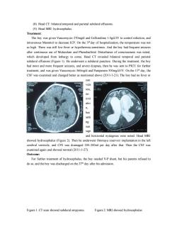
(8) Head CT: bilateral temporal and parietal subdural effusions. (9) Head MRI: hydrocephalus. Treatment: The boy was given Vancomycin 270mg/d and Ceftizedime 1.0g/d IV to control infection, and intravenous Mannitol to decrease ICP. On the 3rd day of hospitalization, the temperature was not so high. There was still low fever or hypothermia sometimes. And the boy had frequent seizures after continuous use of Midazolam and Phenobarbital. Disturbance of consciousness was noted, which developed from lethargy to coma. Head CT revealed bilateral temporal and parietal subdural effusions (Figure 1). He underwent a subdural puncture. During the treatment, the boy had more and more frequent seizures, and severe dyspnea, then he was sent to PICU for further treatment, and was given Vancomycin 360mg/d and Panipenem 500mg/d IV. On the 11th day, the CSF was examined and changed better as mentioned above (2011-1-21). The boy had no fever or con vuls ion, no irrit atio n, but setti ng sun sign and horizontal nystagmus were noted. Head MRI showed hydrocephalus (Figure 2). Then he underwent Ommaya reservoir implantation in the left cerebral ventricle, and CFS was drainaged 200-300ml per day after that. Then the CSF was examined again and showed normal (2011-1-27). Outcome: For further treatment of hydrocephalus, the boy needed V-P shunt, but his parents refused to do so, and the boy was discharged on the 37th day after his admission. Figure 1. CT scan showed subdural empyema Figure 2. MRI showed hydrocephalus
按次数下载不扣除下载券;
注册用户24小时内重复下载只扣除一次;
顺序:VIP每日次数-->可用次数-->下载券;
- 《儿科学》课程作业习题(典型病例)04 iron deficiency anemia.doc
- 《儿科学》课程作业习题(典型病例)03 congenital heart disease-VSD.doc
- 《儿科学》课程作业习题(典型病例)08 diarrhea.doc
- 《儿科学》课程作业习题(典型病例)06 anute nepheritis-1.doc
- 《儿科学》课程作业习题(典型病例)06 nephrotic syndrome-2.doc
- 《儿科学》课程作业习题(复习题)01 questions of children healthcare.doc
- 《儿科学》课程作业习题(复习题)02 questions of neonatal diseases.doc
- 《儿科学》课程作业习题(复习题)05 questions of blood disorders.doc
- 《儿科学》课程作业习题(复习题)04 questions of circulatory system.doc
- 《儿科学》课程作业习题(复习题)03 questions of respiratory diseases.doc
- 《儿科学》课程作业习题(复习题)06 questions of nervous system.doc
- 《儿科学》课程作业习题(复习题)10 questions of infectious diseases.doc
- 《儿科学》课程作业习题(复习题)09 questions of endocrine disorders.doc
- 《儿科学》课程作业习题(复习题)07 questions of urinological system.doc
- 《儿科学》课程作业习题(复习题)08 questions of immune system.doc
- 《儿科学》课程教学资源(授课教案)12 Acute Glomerulonephritis,Nephrotic Syndrome.doc
- 《儿科学》课程教学资源(授课教案)11 Acute Convulsion in Children.doc
- 《儿科学》课程教学资源(授课教案)13 Immunodeficiency.doc
- 《儿科学》课程教学资源(授课教案)10 Nutritional Iron Deficiency Anemia.doc
- 《儿科学》课程教学资源(授课教案)15 Growth Hormone Deficiency.doc
- 《儿科学》课程作业习题(典型病例)07 congenital hypothyroidism.doc
- 《儿科学》课程作业习题(典型病例)03 congenital heart disease-TOF.doc
- 《儿科学》课程作业习题(典型病例)01 ABO incompatibility of neonates.doc
- 《儿科学》课程作业习题(典型病例)02 pneumonia.doc
- 《儿科学》课程作业习题(试卷和答案)双语试卷C卷(试题).doc
- 《儿科学》课程作业习题(试卷和答案)双语试卷B卷(试题).doc
- 《儿科学》课程作业习题(试卷和答案)双语试卷B卷(答案).doc
- 《儿科学》课程作业习题(试卷和答案)双语试卷C卷(答案).doc
- 《儿科学》课程作业习题(试卷和答案)双语试卷A卷(试题).doc
- 《儿科学》课程作业习题(试卷和答案)双语试卷A卷(答案).doc
- 《儿科学》课程教学课件(PPT讲稿)07 新生儿缺氧缺血性脑病 Hypoxic-ischemic Encephalopathy(HIE).pptx
- 《儿科学》课程教学课件(PPT讲稿)06 新生儿败血症 Neonatal Septicemia.pptx
- 《儿科学》课程教学课件(PPT讲稿)21 Chronic Gastritis in Children.pptx
- 《儿科学》课程教学课件(PPT讲稿)20 Toxic Bacillary Dysentery.pptx
- 《儿科学》课程教学课件(PPT讲稿)27 Inflammation Causes Cholesterol Redistribution by Diverting Cholesterol from Circulation to Tissue Tompartments.pptx
- 《儿科学》课程教学课件(PPT讲稿)26 Rotavirus Infection in Children.pptx
- 《儿科学》课程教学课件(PPT讲稿)25 Scarlet Fever.pptx
- 《儿科学》课程教学课件(PPT讲稿)24 Mumps(Epidemic parotitis).pptx
- 《儿科学》课程教学课件(PPT讲稿)23 Infantile Hepatitis Syndrome.pptx
- 《儿科学》课程教学课件(PPT讲稿)22 Infantale Diarrhea and Fluid Therapy.pptx
