山东第一医科大学(泰山医学院):《医学影像学》课程教学资源(授课教案)5.CNS1

Teaching Plan Name:Yu Guang-hui Academic Year:2012-2013 Term:2 Date:2013.3.22 Period:3-4
Teaching Plan Name: Yu Guang-hui Academic Year: 2012-2013 Term: 2 Date: 2013.3.22 Period: 3-4

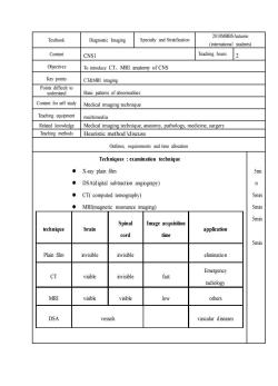
2010MBBSAutumn Textbook Diagnostic Imaging Specialty and Stratification (international students) Content CNS1 Teaching hours2 Objectives To introduce CT,MRI anatomy of CNS Key points CT&MRI imaging PB可 Basic palters of abnormalities Content for self study Medical imaging technique Teaching equipment multimedia Related knowledge Medical imaging technique,anatomy.pathology,medicine.surgery Teaching methods Heuristic method \discuss Outlines requirements and time allocation Techniques examination technique ◆X-ray plain film 5mi DSA(digital subtraction angiograpy) n ●CT(computed tomography) Smin MRI(magnetic resonance imaging) 5min Image acquisition 5min Spinal technique brain application cord time 5min Plain film invisible invisible elimination Emergency CT nvisible fast radiology MRI visible visible low others DSA vessels vascular diseases
Textbook Diagnostic Imaging Specialty and Stratification 2010MBBSAutumn (international students) Content CNS1 Teaching hours 2 Objectives To introduce CT、MRI anatomy of CNS Key points CT&MRI imaging Points difficult to understand Basic patterns of abnormalities Content for self study Medical imaging technique Teaching equipment multimedia Related knowledge Medical imaging technique, anatomy, pathology, medicine, surgery Teaching methods Heuristic method \discuss Outlines, requirements and time allocation Techniques : examination technique ⚫ X-ray plain film ⚫ DSA(digital subtraction angiograpy) ⚫ CT( computed tomography) ⚫ MRI(magnetic resonance imaging) technique brain Spinal cord Image acquisition time application Plain film invisible invisible elimination CT visible invisible fast Emergency radiology MRI visible visible low others DSA vessels vascular diseases 5mi n 5min 5min 5min 5min

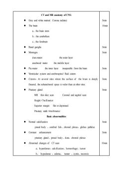
CT and MR anatomy of CNS: Grey and white matter(Corona radiate) 5min ·The brain 10min a、the brain stem b、the cerebellum c、the forebrain ●Basal ganglia 5min ·Meninges 5min dura mater the outer layer arachnoid mater the middle layer ●Pia mater the inner layer inseparable from the brain 5min Ventricular system and cerebrospinal fluid cistem Cistern:At several sites where the surface of the brain is deeply 5min fissured,the subarachnoid space is wider than at other sites. ●Pituitary gland 5min MR thin slice scan Coronal and sagittal scan Height:<7millimeter Superior margin:flat or depressed Pituitary stalk:<4millimeter Basic abnormalities ·Nomal calcification 5min pineal body、cerebral falx、choroid plexus、globus pallidus Contrast enhancement 5min pituitary gland、pineal body、dura、choroid plexus Abnormal changes of CT scan 10min a、hyperdense:calcification、hemorrhage、tumor b、hypodense:edema、tumor、cystis、necrosis
CT and MR anatomy of CNS: ⚫ Grey and white matter( Corona radiate) ⚫ The brain a、the brain stem b、the cerebellum c、the forebrain ⚫ Basal ganglia ⚫ Meninges dura mater the outer layer arachnoid mater the mid dle layer ⚫ Pia mater the inner layer inseparable from the brain ⚫ Ventricular system and cerebrospinal fluid cistern ⚫ Cistern:At several sites where the surface of the brain is deeply fissured, the subarachnoid space is wider than at other sites. ⚫ Pituitary gland MR thin slice scan Coronal and sagittal scan Height:<7millimeter Superior margin: flat or depressed Pituitary stalk:<4millimeter Basic abnormalities ⚫ Normal calcification pineal body 、cerebral falx、choroid plexus、globus pallidus ⚫ Contrast enhancement pituitary gland、pineal body、dura、choroid plexus ⚫ Abnormal changes of CT scan a、hyperdense:calcification、hemorrhage、tumor b、 hypodense :edema、 tumor 、cystis、necrosis 5min 10min 5min 5min 5min 5min 5min 5min 5min 10min

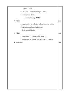
lipoma、fluid c、isodense:chronic hamorhage、tumor dheterogeneous density Abnormal change of MRI ●T1WI: 5min a.hyperintensity:fat or lipoid,melanin,constrast medium b.hypointensity:edema、fluid、tumor fibrosis and calcification ·T2W1: 5min a.hyperintensity:edema,fluid、tumor、 b.hypointensity fibrosis and calcification melanin ●mass effect 5min
lipoma、 fluid c、 isodense :chronic hamorrhage、 tumor d、heterogeneous density Abnormal change of MRI ⚫ T1WI: a. hyperintensity:fat or lipoid、melanin、constrast medium b. hypointensity:edema、fluid、tumor fibrosis and calcification ⚫ T2WI: a. hyperintensity : edema、fluid、tumor 、 b. hypointensity : fibrosis and calcification 、 melanin ⚫ mass effect 5min 5min 5min
按次数下载不扣除下载券;
注册用户24小时内重复下载只扣除一次;
顺序:VIP每日次数-->可用次数-->下载券;
- 山东第一医科大学(泰山医学院):《医学影像学》课程教学资源(授课教案)4.Foundations of Sonagraphy.doc
- 山东第一医科大学(泰山医学院):《医学影像学》课程教学资源(授课教案)3.Radioloy overview 3.doc
- 山东第一医科大学(泰山医学院):《医学影像学》课程教学资源(授课教案)2.Radiolgy overview 2.doc
- 山东第一医科大学(泰山医学院):《医学影像学》课程教学资源(授课教案)1.Radiology overview 1.doc
- 山东第一医科大学(泰山医学院):《医学影像学》课程教学大纲(英文版)Course syllabus-diagnostic imaging.doc
- 《妇产科学》课程教学资源(PPT课件讲稿)产后出血的诊断与处理.ppt
- 《妇产科学》课程教学资源(PPT课件讲稿)营养孕期(妊娠期营养).ppt
- 《妇产科学》课程教学资源(PPT课件讲稿)孕产妇用药.ppt
- 《妇产科学》课程教学资源(PPT课件讲稿)妊娠合并卵巢肿瘤诊疗进展.ppt
- 《妇产科学》课程教学资源(PPT课件讲稿)新生儿复苏策略新进展.ppt
- 《妇产科学》课程教学资源(PPT课件讲稿)孕前和孕期保健指南(第一版).ppt
- 《妇产科学》课程教学资源(PPT课件讲稿)软产道裂伤问题.ppt
- 《妇产科学》课程教学资源(PPT课件讲稿)异位妊娠(安徽中医药高等专科学校).ppt
- 《妇产科学》课程教学资源(PPT课件讲稿)妊娠期高血压疾病的新分类及诊治.ppt
- 《妇产科学》课程教学资源(PPT课件讲稿)外阴上皮内非瘤变 Nonneoplastic Epithelial Disorders of Vulva.ppt
- 《妇产科学》课程教学资源(PPT课件讲稿)盆底功能障碍性及生殖器官损伤疾病.ppt
- 《妇产科学》课程教学资源(PPT课件讲稿)抗凝药物在产科中的应用(二).ppt
- 《妇产科学》课程教学资源(PPT课件讲稿)怀孕和辐射.ppt
- 《妇产科学》课程教学资源(PPT课件讲稿)妊娠滋养细胞疾病(Gestational Trophoblastic Disease, GTD).ppt
- 《妇产科学》课程教学资源(PPT课件讲稿)不孕症 infertility、辅助生殖技术.ppt
- 山东第一医科大学(泰山医学院):《医学影像学》课程教学资源(授课教案)6.CNS2.doc
- 山东第一医科大学(泰山医学院):《医学影像学》课程教学资源(授课教案)7.CNS3.doc
- 山东第一医科大学(泰山医学院):《医学影像学》课程教学资源(授课教案)8.CNS4.doc
- 山东第一医科大学(泰山医学院):《医学影像学》课程教学资源(授课教案)9.Normal Chest X-ray.doc
- 山东第一医科大学(泰山医学院):《医学影像学》课程教学资源(授课教案)10.Thoracic -Basic pathologic changes.doc
- 山东第一医科大学(泰山医学院):《医学影像学》课程教学资源(授课教案)11.Pulmonary inflammatory disease.doc
- 山东第一医科大学(泰山医学院):《医学影像学》课程教学资源(授课教案)12.Pulmonary Tumors.doc
- 山东第一医科大学(泰山医学院):《医学影像学》课程教学资源(授课教案)13.Cardiac imaging.doc
- 山东第一医科大学(泰山医学院):《医学影像学》课程教学资源(授课教案)14.GI_tract-1.doc
- 山东第一医科大学(泰山医学院):《医学影像学》课程教学资源(授课教案)15.GI_tract-2.doc
- 山东第一医科大学(泰山医学院):《医学影像学》课程教学资源(授课教案)16.GI_tract-3.doc
- 山东第一医科大学(泰山医学院):《医学影像学》课程教学资源(授课教案)17.GI_tract-4.doc
- 山东第一医科大学(泰山医学院):《医学影像学》课程教学资源(授课教案)18.Musculoskeletal Imaging1.doc
- 山东第一医科大学(泰山医学院):《医学影像学》课程教学资源(授课教案)19.Musculoskeletal Imaging2.doc
- 山东第一医科大学(泰山医学院):《医学影像学》课程教学资源(授课教案)20..Musculoskeletal Imaging3.doc
- 山东第一医科大学(泰山医学院):《医学影像学》课程教学资源(授课教案)21.Musculoskeletal Imaging4.doc
- 山东第一医科大学(泰山医学院):《医学影像学》课程教学资源(授课教案)22.Genitourinary 1.doc
- 山东第一医科大学(泰山医学院):《医学影像学》课程教学资源(授课教案)23.Genitourinary 2.doc
- 山东第一医科大学(泰山医学院):《医学影像学》课程教学资源(授课教案)24.Genitourinary 3.doc
- 山东第一医科大学(泰山医学院):《医学影像学》课程实验指导 Practical Guide to Diagnostic Imaging.doc
