《病原生物学(人体寄生虫学)》课程实验课件(英文)03 tremode
Human ParasitologyDepartment of Pathogenic and Immunolog)SHIHEZIUNIVERSITY
Human Parasitology Department of Pathogenic and Immunology Department of Pathogenic and Immunology SHIHEZI SHIHEZI UNIVERSITY
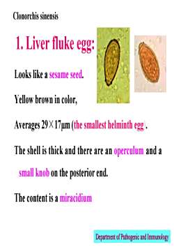
Clonorchis sinensis1. Liver fluke egg:Looks like a sesame seedYellow brown in color,Averages 29 X 17um (the smallest helminth eggThe shellis thick and there are an operculum and asmall knob on the posterior endThe content is a miracidiumDepartment of Pathogenic and Immunology
1. Liver fluke egg: Looks like a sesame seed. Yellow brown in color, Averages 29× 17µm (the smallest helminth egg). The shell is thick and there are an operculum and a small knob on the posterior end. The content is a miracidium Clonorchis sinensis Department of Pathogenic and Immunology Department of Pathogenic and Immunology
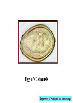
Egg of C. sinensisDepartmentofPathogenicand Immunology
Egg of C. sinensis Department of Pathogenic and Immunology Department of Pathogenic and Immunology
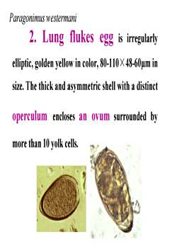
Paragonimus westermani2. Lungg flukes egg is rregularlyelliptic, golden yellow in color, 80-110X 48-60um insize. The thick and asymmetric shell with a distinctoperculum encloses an ovumn surrounded bymore than 10 yolk cells
2. Lung flukes egg is irregularly elliptic, golden yellow in color, 80-110× 48-60µm in size. The thick and asymmetric shell with a distinct operculum encloses an ovum surrounded by more than 10 yolk cells. Paragonimus westermani
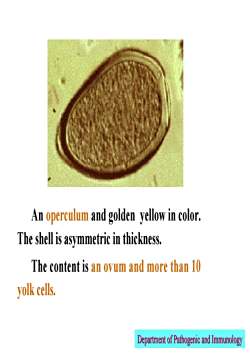
An operculum and golden yellow in color.The shell is asymmetric in thicknessThe content is an ovum and more than 10yolk cells.Department of Pathogenic and Immunology
An operculum and golden yellow in color. The shell is asymmetric in thickness. The content is an ovum and more than 10 yolk cells. Department of Pathogenic and Immunology Department of Pathogenic and Immunology
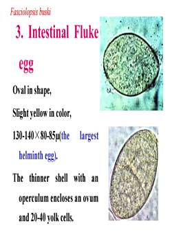
Fasciolopsis buski3. Intestinal FlukeeggOval in shape,Slight yellow in color,130-140 X 80-85μ(thelargesthelminth egg)The thinner shell with anoperculum encloses an ovumand 20-40 yolk cells
3. Intestinal Fluke egg Oval in shape, Slight yellow in color, 130-140× 80-85µ(the largest helminth egg). The thinner shell with an operculum encloses an ovum and 20-40 yolk cells. Fasciolopsis buski
4. Schistosoma eggOval in shape, slight yellow in color, 89 x 67umshell is thin without an operculum but with a lateralspine.The content is a miracidiumUnder the electron microscope there are manymicro-tubules on the shell, through which the solubleegg antigen (SEA) secreted by a miracidium.Department of Pathogenic and Immunology
4. Schistosoma egg Oval in shape, slight yellow in color, 89 x 67µm, shell is thin without an operculum but with a lateral spine. The content is a miracidium. Under the electron microscope there are many micro-tubules on the shell, through which the soluble egg antigen (SEA) secreted by a miracidium. Department of Pathogenic and Immunology Department of Pathogenic and Immunology
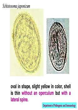
Schistosoma japonicumoval in shape, slight yellow in color, shelis thin without an operculum but with alateralspineDepartment of Pathogenic and Immunology
oval in shape, slight yellow in color, shell is thin without an operculum but with a lateral spine. Schistosoma japonicum Department of Pathogenic and Immunology Department of Pathogenic and Immunology
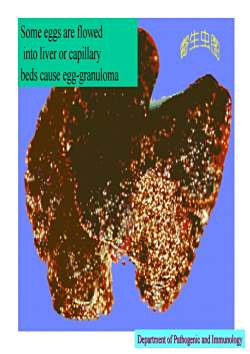
Some eggs are flowed新生虫肉into liver or capillarybeds cause egg-granulomaDepartment of Pathogenicand Immunology
Some eggs are flowed into liver or capillary beds cause egg-granuloma Department of Pathogenic and Immunology Department of Pathogenic and Immunology
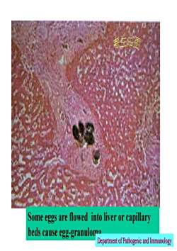
生虫Some eggs are flowed into liver or capillarybeds cause egg-granulomaDepartment of Pathogenic and Immunology
Some eggs are flowed into liver or capillary beds cause egg-granulomaDepartment of Pathogenic and Immunology Department of Pathogenic and Immunology
按次数下载不扣除下载券;
注册用户24小时内重复下载只扣除一次;
顺序:VIP每日次数-->可用次数-->下载券;
- 《病原生物学(人体寄生虫学)》课程教学课件(讲稿)02 丝虫、旋毛虫.pdf
- 《病原生物学(人体寄生虫学)》课程教学课件(讲稿)03 猪牛带绦虫.pdf
- 《病原生物学(人体寄生虫学)》课程教学课件(讲稿)01 蛔钩蛲虫幻灯.pdf
- 《病原生物学(人体寄生虫学)》课程教学课件(讲稿)04 包生绦虫 短小膜壳绦虫 裂头绦虫.pdf
- 《病原生物学(人体寄生虫学)》课程教学课件(讲稿)07 概述 阿米巴 兰氏贾第鞭毛虫.pdf
- 《病原生物学(人体寄生虫学)》课程教学课件(讲稿)05 肝、肺吸虫姜片虫.pdf
- 《病原生物学(人体寄生虫学)》课程教学课件(讲稿)06 血吸虫.pdf
- 《病原生物学(人体寄生虫学)》课程教学课件(讲稿)08 人体疟原虫.pdf
- 《病原生物学(人体寄生虫学)》课程教学课件(讲稿)13 蚤虱蛉.pdf
- 《病原生物学(人体寄生虫学)》课程教学课件(讲稿)09 利什曼原虫.pdf
- 《病原生物学(人体寄生虫学)》课程教学课件(讲稿)12 医学节肢动物绪论、蚊、蝇.pdf
- 《病原生物学(人体寄生虫学)》课程教学课件(讲稿)14 蜱螨.pdf
- 《病原生物学(人体寄生虫学)》课程教学课件(讲稿)11 机会致病性原虫.pdf
- 《医学免疫学》课程教学课件(2017讲稿)第2章 免疫器官和组织.pdf
- 《医学免疫学》课程教学课件(2017讲稿)第1章 免疫学概论.pdf
- 《医学免疫学》课程教学课件(2017讲稿)第4章 抗体.pdf
- 《医学免疫学》课程教学课件(2017讲稿)第5章 补体系统.pdf
- 《医学免疫学》课程教学课件(2017讲稿)第3章 抗原.pdf
- 《医学免疫学》课程教学课件(2017讲稿)第6章 细胞因子.pdf
- 《医学免疫学》课程教学课件(2017讲稿)第12章 T细胞介导的细胞免疫应答.pdf
- 《病原生物学(人体寄生虫学)》课程实验课件(英文)04 Ascaris Lumbricoides and Trichuris trichiura.pdf
- 《病原生物学(人体寄生虫学)》课程实验课件(英文)05 Nematodes.pdf
- 《病原生物学(人体寄生虫学)》课程实验课件(英文)02 cestoda.pdf
- 《病原生物学(人体寄生虫学)》课程实验课件(英文)01 protozo.pdf
- 《病原生物学(人体寄生虫学)》课程教学课件(2012)丝虫、旋毛虫.pdf
- 《病原生物学(人体寄生虫学)》课程教学课件(2012)医学节肢动物 蜱螨.pdf
- 《病原生物学(人体寄生虫学)》课程教学课件(2012)医学节肢动物 昆虫纲(下).pdf
- 《病原生物学(人体寄生虫学)》课程教学课件(2012)绦虫概论、猪带绦虫、牛带绦虫.pdf
- 《病原生物学(人体寄生虫学)》课程教学课件(2012)医学节肢动物 昆虫纲(上).pdf
- 《病原生物学(人体寄生虫学)》课程教学课件(2012)细粒棘球绦虫、多房棘球绦虫、微小膜壳绦虫.pdf
- 《病原生物学(人体寄生虫学)》课程教学课件(2012)肺吸虫、血吸虫.pdf
- 《病原生物学(人体寄生虫学)》课程教学课件(2012)蛲虫、钩虫.pdf
- 《病原生物学(人体寄生虫学)》课程教学课件(2012)蛔虫、鞭虫.pdf
- 《病原生物学(人体寄生虫学)》课程教学课件(2012)吸虫概论、肝、肠吸虫.pdf
- 《病原生物学(人体寄生虫学)》课程教学课件(2012)疟原虫-2/2.pdf
- 《病原生物学(人体寄生虫学)》课程教学课件(2012)疟原虫-1/2.pdf
- 《病原生物学(人体寄生虫学)》课程教学课件(2012)机会性致病原虫.pdf
- 《病原生物学(人体寄生虫学)》课程教学课件(2012)原虫——鞭毛虫.pdf
- 《病原生物学(人体寄生虫学)》课程教学课件(2012)人体寄生虫学总论.pdf
- 《病原生物学(人体寄生虫学)》课程教学课件(2012)原虫概论及阿米巴原虫.pdf
